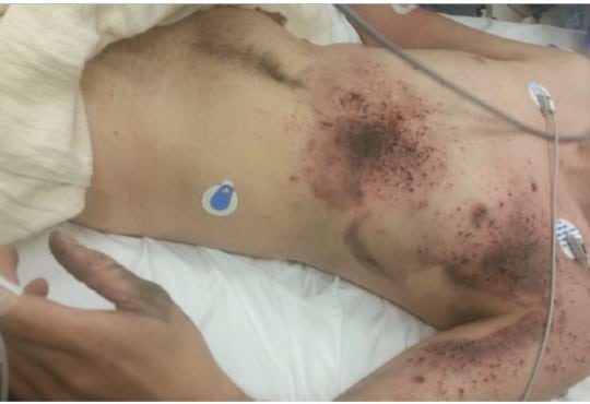
It is important to rapidly diagnosis and treat rhabdomyolysis in order to decrease morbidity and mortality. To date there are no reports in the emergency medicine literature on the use of point-of-care ultrasound in the diagnosis of rhabdomyolysis.
Electronic cigarettes (also known as e-cigarettes or e-cigs) are becoming a popular method of recreational nicotine use over recent years. The growth of new brands and devices has been outpacing the FDA’s ability to regulate them. As a result, some of these devices fail without warning, most likely from malfunction of the lithium-ion batteries that are in close proximity to volatile compounds within the device.
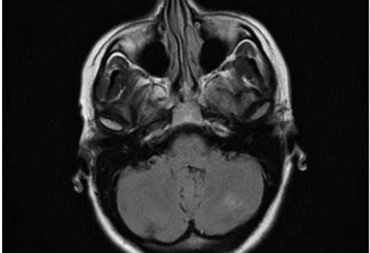
Unruptured posterior communicating artery (PCOM) aneurysms can be difficult to diagnose and, when large (≥ 7mm), represent a substantial risk to the patient. While most unruptured PCOM aneurysms are asymptomatic, when symptoms do occur, clinical manifestations typically include severe headache (HA), visual acuity loss, and cranial nerve deficit. This case report describes an atypical initial presentation of a large unruptured PCOM aneurysm with symptoms mimicking trigeminal neuralgia, without other associated cranial nerve palsies or neurologic deficits.
Spermatic cord anesthesia block (SCAB) is a useful technique for providing anesthesia to males with scrotal pain. This technique has been described and published in the urology and anesthesia literature for more than 40 years. Initially described as a blind injection, anesthesia of the spermatic cord provides pain control to the scrotal contents. The technique can easily be performed under ultrasound guidance by emergency physicians and should be considered a useful option when seeking to provide pain relief to male patients with scrotal pain.
Two Cases of Anti-NMDA Receptor Encephalitis
Jesse Baker, BA, et al.
Anti-N-methyl-D-aspartate receptor (anti-NMDAR) encephalitis is a form of autoimmune encephalitis with prominent neuropsychiatric features. Patients present with acute psychosis, memory impairment, dyskinesias, seizures, and/or speech disorders. The clinical course is often complicated by respiratory failure, requiring intubation. Approximately half of patients are found to have an associated ovarian tumor, which expresses NMDAR. Recognition of anti-NMDAR encephalitis by emergency physicians is essential in order to initiate early treatment and avoid psychiatric misdiagnosis. The disease is highly treatable with tumor removal and immunosuppression, and most patients demonstrate a full recovery. In this case series, we report two cases of anti-NMDAR encephalitis in adult women in the United States and provide a review of the literature.
A Rare but Important Clinical Presentation of Induced Methemoglobinemia
Faried Banimahd, MD et al.
Phenazopyridine is considered one of the classic causes of drug-induced methemoglobinemia. It is often taught as such and seen in board review courses. Nevertheless, the epidemiology is unknown, presentation quite rare, and less than five cases have been reported in PubMed in over 35 years.1-4 We present a case with a different set of patient characteristics than seen in the few recent case reports, and an approach to treatment that validates further uniqueness, justifying reporting the case in the literature. In particular, the patient was a young otherwise-healthy adult, with the initial diagnosis and decision to treat based on clinical grounds versus laboratory values.
A Curious Case of Right Upper Quadrant Abdominal Pain
Andrew Grock, MD et al.
An otherwise healthy 36-year-old man presented with sudden-onset right upper quadrant abdominal pain and vomiting. A bedside ultrasound, performed to evaluate hepatobiliary pathology, revealed a normal gallbladder but free intraperitoneal fluid. After an expedited CT and emergent explorative laparotomy, the patient was diagnosed with a small bowel obstruction with ischemia secondary to midgut volvulus. Though midgut volvulus is rare in adults, delays in definitive diagnosis and management can result in bowel necrosis. Importantly, an emergency physician must be able to recognize bedside ultrasound findings associated with acutely dangerous intrabdominal pathology.
Volume 17, Issue 4, July 2016
Siamak Moayedi, MD, et al.
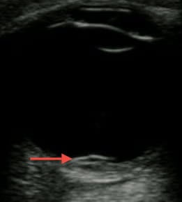
A three-year-old girl presented to the emergency department (ED) for five days of pain and decreased mobility of the left shoulder. She had been evaluated in the ED five days prior for shoulder pain after a minor slip and fall with negative clavicle radiographs, and was discharged home with supportive care. Since the initial visit, her shoulder pain increased and she would not use her arm. Physical examination demonstrated subtle swelling of the left anterior shoulder without erythema, warmth, or fluctuance. Her exam yielded mild tenderness to palpation and markedly decreased range of motion secondary to pain. Point-of-care shoulder ultrasound revealed an enlarged deltoid muscle with a heterogeneous fluid collection within the muscle, but no joint effusion (Video).
Volume 17, Issue 4, July 2016
Kelly Kesler, MD, et al.
A 29-year-old female with no significant past medical history presented with palpitations, nausea, diaphoresis and lightheadedness. Symptoms began 15 minutes prior to arrival. She reported several similar episodes previously that self-resolved within seconds, but had no previous medical evaluations for these symptoms. Initial vital signs were significant for blood pressure of 93/61, irregular heart rate between 180 and 200, respiratory rate of 18, and oxygen saturation of 99% on room air. Physical examination was otherwise unremarkable. The electrocardiogram (ECG) is shown in Figure 1. This was interpreted as atrial fibrillation with rapid ventricular rate, and the patient was treated with rate control with no effect. The patient later spontaneously converted to normal sinus rhythm and repeat ECG was notable for delta waves concerning for Wolff-Parkinson-White Syndrome (WPW) as seen in Figure 2. She was admitted to cardiology for cardiac ablation.
Volume 17, Issue 4, June 2016
Zachary Dezman, MD, MS, et al.
A sexually active 35-year old woman presented to the emergency department with intermittent vaginal spotting and pelvic cramping over the preceding four weeks. She had an intrauterine device (IUD) placed three months prior and has never been pregnant. The threads of the IUD and a small amount of blood coming from the cervix were seen on pelvic exam. Laboratory testing revealed a β-human chorionic gonadotropin level of 70,000 mIU/mL. Pelvic ultrasound imaging showed the IUD and a viable intrauterine pregnancy.
Volume 17, Issue 4, June 2016
Monica Lee, MD, et al.
This case describes an emergency department (ED) presentation of ocular syphilis in a human immunodeficiency virus (HIV) infected patient. This is an unusual presentation of syphilis and one that emergency physicians should be aware of. The prevalence of syphilis has reached epidemic proportions since 2001 with occurrences primarily among men who have sex with men (MSM). This is a case of a 24-year-old male who presented to our ED with bilateral painless vision loss. The patient’s history and ED workup were notable for MSM, positive rapid plasmin reagin (RPR) and HIV tests and fundus exam consistent with ocular syphilis, specifically uveitis. Ocular manifestations of syphilis can present at any stage of syphilis. The 2010 Centers for Disease Control and Prevention guidelines now recommend that ocular syphilis be treated as neurosyphilis regardless of the lumbar puncture results. There is a paucity of emergency medicine literature on ocular syphilis. For emergency physicians it is important to be aware of iritis, uveitis, or chorioretinitis as ocular manifestations of neurosyphilis especially in this high-risk population and to obtain RPR and HIV tests in the ED to facilitate early diagnosis, and treatment and to prevent irreversible vision loss.
Volume 17, Issue 3, May 2016
Daniel R. Lasoff, MD et al.
Anti-N-Methyl-D-Aspartate Receptor (NMDAR) Encephalitis is a novel disease discovered within
the past 10 years. Antibodies directed at the NMDAR cause the patient to develop a characteristic
syndrome of neuropsychiatric symptoms. Patients go on to develop autonomic dysregulation and
often have prolonged hospitalizations and intensive care unit stays. There is little literature in
the emergency medicine community regarding this disease process, so we report on a case we
encountered in our emergency department to help raise awareness of this disease process.
Volume 17, Issue 3, May 2016
R. Mason Curtis, MD et al.
Introduction: Upper airway angioedema is a life-threatening emergency department (ED)
presentation with increasing incidence. Angiotensin-converting enzyme inhibitor induced
eScholarship provides open access, scholarly publishing
services to the University of California and delivers a dynamic
research platform to scholars worldwide.
angioedema (AAE) is a non-mast cell mediated etiology of angioedema. Accurate diagnosis by
clinical examination can optimize patient management and reduce morbidity from inappropriate
treatment with epinephrine. The aim of this study is to describe the incidence of angioedema
subtypes and the management of AAE. We evaluate the appropriateness of treatments and
highlight preventable iatrogenic morbidity.
Methods: We conducted a retrospective chart review of consecutive angioedema patients
presenting to two tertiary care EDs between July 2007 and March 2012.
Results: Of 1,702 medical records screened, 527 were included. The cause of angioedema
was identified in 48.8% (n=257) of cases. The most common identifiable etiology was AAE
(33.1%, n=85), with a 60.0% male predominance. The most common AAE management strategies
included diphenhydramine (63.5%, n=54), corticosteroids (50.6%, n=43) and ranitidine (31.8%,
n=27). Epinephrine was administered in 21.2% (n=18) of AAE patients, five of whom received
repeated doses. Four AAE patients required admission (4.7%) and one required endotracheal
intubation. Epinephrine induced morbidity in two patients, causing myocardial ischemia or
dysrhythmia shortly after administration.
Conclusion: AAE is the most common identifiable etiology of angioedema and can be accurately
diagnosed by physical examination. It is easily confused with anaphylaxis and mismanaged with
antihistamines, corticosteroids and epinephrine. There is little physiologic rationale for epinephrine
use in AAE and much risk. Improved clinical differentiation of mast cell and non-mast cell mediated
angioedema can optimize patient management.
Volume 17, Issue 2, March 2016.
Siri Shastry, MD, et al.
Electronic vapor cigarettes (E-cigarettes) were created
in 2003 as an alternative to traditional tobacco cigarettes.
E-cigarettes have been available in the United States since
2006. The typical E-cigarette consists of a cartridge that
contains liquid, an atomizer that heats the liquid (i.e. acts
as a vaporizer), as well as a battery. The liquid contained
within the cartridge contains nicotine, propylene glycol and/
or glycerol as well as flavorings.The consumer uses an
E-cigarette through either pushing a button or inhalation,
which triggers heating and therefore aerosolizes the liquid
within the cartridge, emulating cigarette “smoke.” The newest
E-cigarettes are larger than nicotine cigarettes and employ
stronger, rechargeable batteries as a power source.
Volume 17, Issue 2, March 2016.
Trevonne M. Thompson, MD, et al.
Methylsalicylate-containing rubefacients have been reported to cause salicylate poisoning after
ingestion, topical application to abnormal skin, and inappropriate topical application to normal
skin. Many over-the-counter products contain methylsalicylate. Topical salicylates rarely produce
systemic toxicity when used appropriately; however, methylsaliclyate can be absorbed through intact
skin. Scrotal skin can have up to 40-fold greater absorption compared to other dermal regions. We
report a unique case of salicylate poisoning resulting from the use of a methylsalicylate-containing
rubefacient to facilitate masturbation in a male teenager. Saliclyate toxicity has not previously been
reported from the genital exposure to methylsaliclyate.
Volume 17, Issue 2, March 2016.
Timothy D. Roberts, MBChB
An eight-year-old boy presented to the emergency
department (ED) with a 2cm-long laceration over the
prepatellar region of his left knee after falling over and
cutting his knee on broken glass. Physical examination
demonstrated the laceration breached the dermis but
otherwise there was no obvious defect in the deep fascial
layer.
Volume 17, Issue 2, March 2016.
Kristin H. Dwyer, MD, MPH, et al.
A 26-year-old female presented to the emergency
department with three days of subjective fevers, dry cough
and pleuritic chest discomfort. On exam, her vital signs
were significant for a heart rate of 106/minute and oxygen
saturation of 95% on room air. Her lung exam revealed
decreased breath sounds at the right base. A bedside lung
ultrasound and a chest radiograph were performed.
Volume 17, Issue 2, March 2016.
Meina J. Michael, BS, et al.
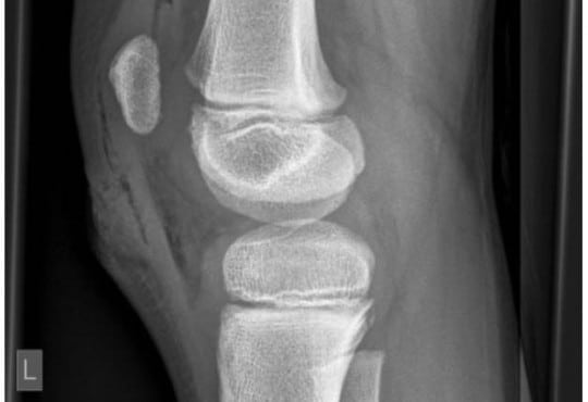
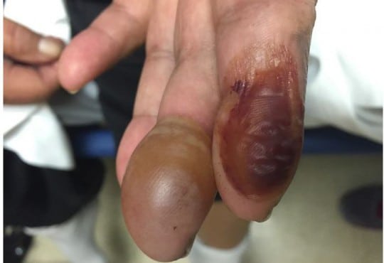
A 63-year-old female with insulin-dependent type II
diabetes mellitus and end-stage renal disease presented to the
emergency department with spontaneous blistering to the tips
of her left index and middle fingers. The blisters had gradually
become tense and mildly painful over the preceding 10 days.
She denied burn injury, trauma, fever, or new medications.
On physical exam, the patient was noted to have a tense,
nontender bullae on the pad of the left middle finger, and a
collapsed, hemorrhagic bullae on the left index finger. There
were no signs of inflammation or infection. A radiograph of
the left hand, complete blood count, and basic metabolic panel
were unremarkable. The diagnosis of bullosis diabeticorum
was made, and supported by a consulting endocrinologist.
Volume 17, Issue 2, March 2016.
Samuel L. Burleson, MD, et al.
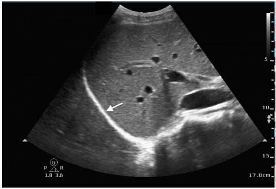
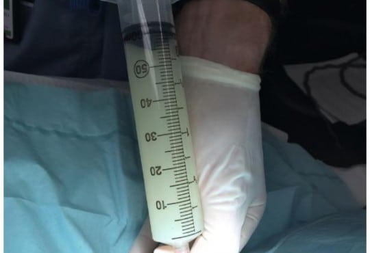
A 58-year-old female with a past medical history of
hepatitis C virus-induced cirrhosis presented to the emergency
department with three days of increasing abdominal pain,
chills, and nausea and vomiting. Abdominal physical
examination revealed gross ascites with fluid wave. Diagnostic
paracentesis resulted in the aspiration of approximately 60mL
of white turbid peritoneal fluid (Figure).
Volume 17, Issue 1, January 2016.
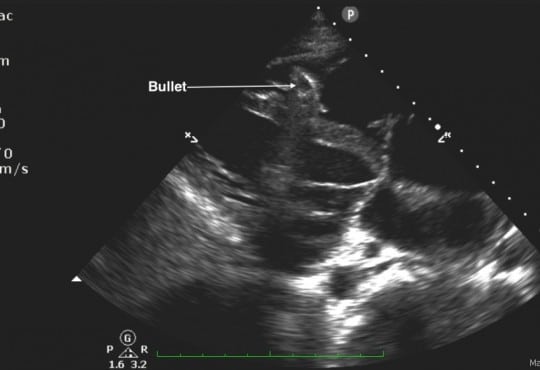
Abdullah Bakhsh, MD, et al.
A 25-year-old male was brought in by ambulance to
the emergency department (ED) after sustaining a gunshot
wound to his chin and left shoulder. Upon arrival to the
ED, his airway was intact without evidence of blood in the
oropharynx. He was found to have slightly diminished breath
sounds on the left side, with respirations at 34 breaths per
minute, a blood pressure of 72/50mmHg, and a heart rate of
76 beats per minute with cool extremities and poor peripheral
pulses. His focused abdominal sonography in trauma exam
showed a foreign body within the right ventricle without a
pericardial effusion (Figure 1 and Video). An upright portable
chest radiograph performed immediately thereafter showed
blunting of the left costophrenic angle with a bullet fragment
overlying the cardiac shadow (Figure 2).
Volume 17, Issue 1, January 2016.
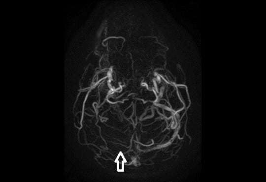
Rohat Ak, MD, et al.
A 45-year-old man presented with headache for two days.
He described the quality of headache as throbbing, and it was
unilateral. There was no history of fever, vomiting, blurred
vision, ear discharge or trauma, no relevant past medical or
drug history and no family history of note. On examination,
he was afebrile with pulse 76/min, regular, blood pressure of
130/80mmHg. His pupils and speech appeared normal. There
were no papilledema, sensory deficit, focal neurological deficit
or signs of meningeal irritation. Hyperdensity of right transverse
sinus (Figure 1) and superior sagittal sinus was identified on
unenhanced computed tomography (CT). Magnetic resonance
venography (MRV) demonstrated lack of flow in right transverse
sinus (Figure 2) and superior sagittal sinus.
Volume 17, Issue 1, January 2016.
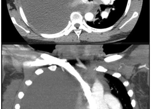
AnnaKate Deal, MD, et al.
We present the case of a 34-year-old woman presenting to
the emergency department (ED) with dyspnea, cough, and fever.
She was found to have a tension hydrothorax and was treated
with ultrasound-guided thoracentesis in the ED. Subsequent
inpatient evaluation showed the patient had disseminated
endometriosis. Tension hydrothorax has not been previously
described in the literature as a complication of this disease.
Volume 16, Issue 7, December 2015.
Tara Murphy, et al.
Posterior reversible encephalopathy syndrome (PRES) is an unusual condition typified by acute
visual impairment caused by sudden, marked parieto-occipital vasogenic edema. Thought to be
inflammatory in origin, it has been described in patients undergoing chemotherapy, with autoimmune
disease, and in some infections. We report a case of PRES that occurred one week after an episode
of acute pancreatitis in an otherwise healthy 40-year-old female. There was progressive visual
impairment over a 24-hour period with almost complete visual loss, with characteristic findings on
magnetic resonance imaging. After treatment with steroids, the visual loss recovered. Clinicians
should retain an index of suspicion of this rare condition in patients with visual impairment after acute
pancreatitis.
Volume 16, Issue 7, December 2015.
Jesse Z. Kellar, MD, et al.
A 14-year-old African American male presented to the emergency department with worsening left
eye redness, swelling, and vision loss over the preceding three days. History was notable for similar
eye redness and swelling without vision loss four months earlier, which improved following a brief
course of prednisone. He endorsed mild eye irritation and tearing with bright lights. There was
no history of fever, respiratory symptoms or trauma. Mother was medicating patient with leftover
antibiotic eye drops x3 days without improvement. Physical examination on presentation notable
for proptosis of left eye, lid, and periorbital swelling, mild scleral injection, and central vision loss in
affected eye (20/200 OS, 20/25 OD). Extraocular movements and pupillary exam were normal. No
corneal fluorescein uptake, abnormal cell, flare, or siedel sign were seen during slit lamp exam. Eye
pressures were 24 mmHg in both eyes. Bedside ultrasonography was performed (Figure 1 showing
retinal detachment, Ultrasound Video 2 showing detachment in orbital scan).
Volume 16, Issue 7, December 2015.
Laura J. Fil, DO, et al.
Multiple sclerosis (MS) is an immune mediated inflammatory disease that attacks myelinated axons
in the central nervous system. Dalfampridine (4-aminopyridine) was approved by the Food and
Drug Administration in January 2010 for treatment of MS. Our patient was a 34-year-old male with a
history of MS, who was brought to the emergency department after being found unresponsive. His
current medications were valacyclovir, temazepam, dalfampridine (4-AP) and a tysabri intravenous
(IV) infusion. Fifteen minutes after arrival the patient seized. The seizures were refractory to
benzodiazepines, barbiturates and phenytoin. The 4-AP level was 530ng/mL (25ng/mL and 49ng/
mL). The patient stopped seizing on hospital day 3 and was discharged 14 days later with normal
mental status and neurologic exam. 4-AP is a potassium channel blocker that blocks the potassium
ion current of repolarization following an action potential. The blockade of the potassium channel at
the level of the membrane widens the action potential and enhances the release of acetylcholine,
thus increasing post-synaptic action potentials. The treatment of patients with 4-AP overdose is
supportive. Animal data suggest that patients with toxic levels of 4-AP may respond to phenytoin.
Our case illustrates the highest recorded level of 4-AP in an overdose. Our patient appeared to be
refractory to a combination of high doses of anticonvulsants and only improved with time.
Volume 16, Issue 7, December 2015.
Erik A. Berg, MD
A 60-year-old female with a history of
ventriculoperitoneal shunt (VPS) placement three years
prior presented with a painful abdominal wall mass.
The patient denied fevers, nausea, vomiting, headaches,
or dizziness. Physical exam revealed an afebrile, wellappearing
female with a raised, erythematous, fluctuant mass
on the right lower abdominal wall. She had no abdominal
tenderness otherwise. Labs were unremarkable. A bedside
ultrasound revealed a complex fluid collection over the
area of fluctuance that tracked along the course of the VPS
tubing into the abdomen. Plan for incision and drainage was
deferred. Neurosurgery was consulted. The neurosurgeon
attempted to tap the shunt but encountered very high
resistance. The patient was admitted for intravenous antibiotics for VPS infection and malfunction.