| Author | Affiliation |
|---|---|
| Timothy D. Roberts, MBChB | Starship Children’s Hospital, Department of Paediatric Orthopaedics, Auckland, New Zealand |
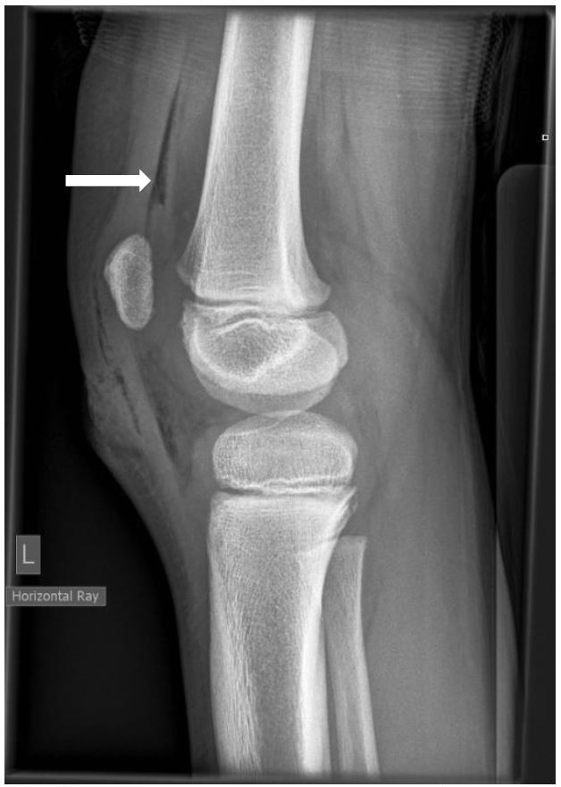
An eight-year-old boy presented to the emergency department (ED) with a 2cm-long laceration over the prepatellar region of his left knee after falling over and cutting his knee on broken glass. Physical examination demonstrated the laceration breached the dermis but otherwise there was no obvious defect in the deep fascial layer. He had a free range of motion of his knee and clinically his extensor mechanism was intact; however, a plain lateral radiograph showed that he had pneumarthrosis of his knee joint. Within six hours of injury the wound was formally explored in the operating room and a small breach in the knee capsule was found. The wound edges were debrided, the knee joint irrigated and the skin closed primarily. Following surgery he received 24 hours of antibiotic coverage with a first-generation cephalosporin.
Gas in the joint or “pneumarthrosis” in the context of trauma is not uncommon around superficial joints such as the knee and indicates that there is a penetrating wound with intra-articular extension.1 This implies that the joint has been inoculated with bacteria and pyoarthrosis is possible if not managed appropriately.1 The standard treatment for a traumatic arthrotomy is antibiotic coverage and prompt surgical debridement in the operating room with lavage of the soft tissues and joint.2 In the absence of gross contamination primary closure can be performed.2
Even innocuous-looking lacerations adjacent to or overlying superficial joints can lead to a traumatic arthrotomy. Plain radiographs should be obtained and a high suspicion for intra-articular extension maintained. For lacerations around superficial joints there should be a low threshold for exploring and washing these wounds out in the operating room rather than closing primarily in the ED.
Footnotes
Section Editor: Rick A. McPheeters, DO
Full text available through open access at http://escholarship.org/uc/uciem_westjem
Address for Correspondence: Timothy D. Roberts, MBChB, Starship Children’s Hospital, 2 Park Road, Grafton, Auckland 1023, New Zealand. Email: timroberts.nz@gmail.com. 3 / 2016; 17:184 – 185
Submission history: Revision received November 18, 2015; Submitted December 21, 2015; Accepted December 22, 2015
Conflicts of Interest: By the WestJEM article submission agreement, all authors are required to disclose all affiliations, funding sources and financial or management relationships that could be perceived as potential sources of bias. The authors disclosed none.
REFERENCES
1. Nord RM, Quach T, Walsh M, et al. Detection of traumatic arthrotomy of the knee using the saline solution load test. J Bone Joint Surg Am. 2009;91(1):66-70.
2. Konda SR, Davidovitch RI, Egol KA. Open knee joint injuries an evidence-based approach to management. Bull Hosp Jt Dis. 2014;72(1):61-9.