
| Author | Affiliation |
|---|---|
| Rahul Sharma, MD, MBA | New York Presbyterian-Weill Cornell Medical Center, Department of Emergency Medicine, New York, New York |
| Hana Yoshikawa, RPA-C | New York Presbyterian-Weill Cornell Medical Center, Department of Emergency Medicine, New York, New York |
| Josyann Abisaab, MD | New York Presbyterian-Weill Cornell Medical Center, Department of Emergency Medicine, New York, New York |
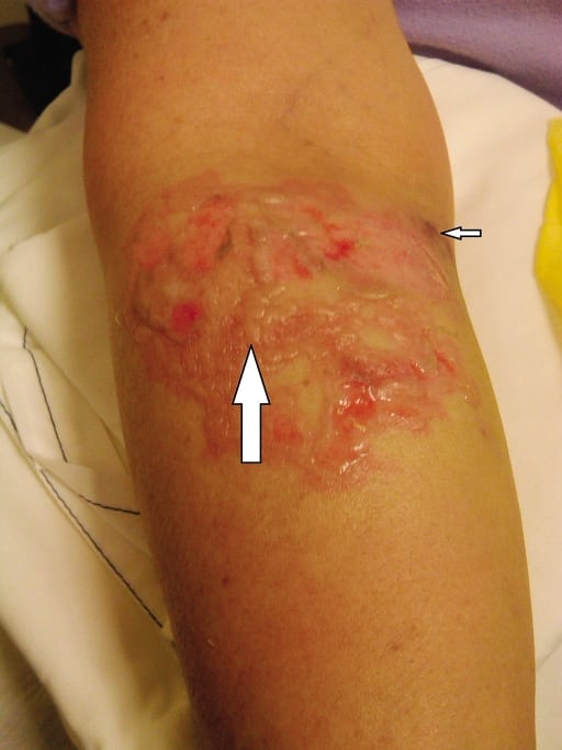
A 58-year-old female presented to the emergency department (ED) with pain and swelling to the right arm after receiving propofol during an outpatient procedure for nasal polyps. During the procedure, it was noted that propofol had infiltrated at the antecubital intravenous (IV) site. A new IV line was inserted, and the procedure was completed. The patient was discharged from the hospital and then presented to our ED.
Examination showed a 7-cm-by-8-cm denuded area with erythema and edema in the antecubital region (1% total body surface area). An hour after presentation, ecchymosis developed medially (Figure). There was small amount of serous drainage and tenderness to palpation. She was neurovascularly intact.
The burn service was consulted. The patient was admitted to the burn unit and treated with antibiotics. She received a skin graft and discharged on post-op day 5 without any complications.
DISCUSSION
Propofol is a widely used anesthetic with many favorable properties, including short half-life, neutral pH, and isotonicity.1 Owing to these factors, extravasation injuries due to propofol are relatively rare, though cases of tissue necrosis have been reported.1–3 Risk factors for injury include cytotoxicity of the solution, infusion pressure, regional anatomical peculiarities, and other patient factors, such as preexisting cutaneous or vascular pathophysiology.4
When extravasation occurs, the infusion must be stopped immediately. If possible, the extravasated fluid should be aspirated before withdrawing the needle, and consider flushing with Ringer’s solution or normal saline.1,2,4 Immediate surgical consultation should be obtained. The risk for tissue damage after extravasation is often underestimated, resulting in potentially limb-threatening morbidity.4
Footnotes
Supervising Section Editor: Sean Henderson, MD
Submission history: Submitted May 29, 2011; Accepted June 10, 2011
Reprints available through open access at http://escholarship.org/uc/uciem_westjem
DOI: 10.5811/westjem.2011.6.6813
Address for Correspondence: Rahul Sharma, MD, MBA
New York Presbyterian-Weill Cornell Medical Center, Department of Emergency Medicine, 525 E 68th St, Rm M-130, New York, NY 10065
E-mail: rahul_sharma@hotmail.com
Conflicts of Interest: By the WestJEM article submission agreement, all authors are required to disclose all affiliations, funding, sources, and financial or management relationships that could be perceived as potential sources of bias. The authors disclosed none.
REFERENCES
1. Roth W, Stephan E, Gardetto A, et al. Extravasation of propofol is associated with tissue necrosis in small children. Pediat Anesth. 2006;16:887–889. [PubMed]
2. Huijpers EJM, Baars JW, Schutte PFE, et al. Propofol extravsation in a breast cancer patient. J Oncol Pharm Practice. 2008;14:195–198. [PubMed]
3. Tokumine J, Sugahara K, Tomori T, et al. Tissue necrosis caused by extravasated propofol. J Anesthesia. 2002;16:358–359. [PubMed]
4. Schummer W, Schummer C, Bayer O, et al. Extravasation injuries in the perioperative setting.Anesth Analg. 2006;100:722–727. [PubMed]