
| Author | Affiliation |
| Steven G. Schauer, DO | Bayne-Jones Army Community Hospital, Department of Emergency Medicine, Fort Polk, Louisiana |
| Justin C. Eisenman, DO | Blanchfield Army Community Hospital, Fort Campbell, Kentucky |
ABSTRACT
A 55-year-old female presented to the emergency department at a small community hospital with cough, fever, dyspnea and blood-streaked sputum. A chest radiograph was ordered. She was diagnosed with pneumonia and discharged home with antibiotics. She returned three days later, afebrile, with worsening dyspnea and gross hemoptysis. She was found to have a murmur reported as chronic but had never been evaluated by echocardiography. A computed tomography chest and echocardiography were performed (Figure). She was diagnosed with a left atrial myxoma (Video). She was transferred and underwent tumor excision.
DISCUSSION
Primary cardiac tumors are rare with an incidence of 0.05%.1 The overwhelming majority of cardiac myxomas are located in the left atrium, followed by the right atrium and then in the ventricles.2,3 Like most cardiac myxomas, the histology in this presentation is benign. The myxoma triad consists of obstructive symptoms (heart failure, shortness of breath), malaise, and embolic events. Physical exam may reveal a murmur known as “tumor plop” that often mimics mitral stenosis.4 The diagnosis is often made when evaluating for etiologies of similar presentation. Echocardiography, chest computed tomography (CT), cardiac magnetic resonance imaging (MRI), and cardiac angiography have all been described. Echocardiography has the advantage of being able to evaluate size, shape, location and mobility in a dynamic fashion with reported sensitivities of 95% and nearly 100% for transthoracic and transesophageal, respectively.5 MRI and CT appear to have lesser reliability when determining tumor origin.6 The prognosis is good with a 96% 10-year survival rate.3
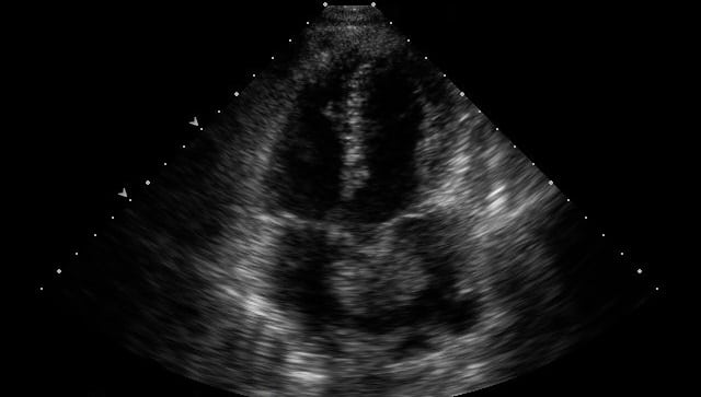
Figure. Echocardiography of patient diagnosed with left atrial myxoma.
Video. Video demonstrates the dynamic left atrial myxoma denoted by arrow.
ACKNOWLEDGEMENTS
Footnotes
Supervising Section Editor: Sean O. Henderson, MD
Full text available through open access at http://escholarship.org/uc/uciem_westjem
Address for Correspondence: Steven G. Schauer, DO, Bayne-Jones Army Community Hospital, 1585 3rd Street, Fort Polk, LA 71459. Email: sgschauer@gmail.com.
Submission history: Submitted September 7, 2014; Accepted September 23, 2014
Conflicts of Interest: By the WestJEM article submission agreement, all authors are required to disclose all affiliations, funding sources and financial or management relationships that could be perceived as potential sources of bias. The authors disclosed none.
REFERENCES
- Nicholls GM, Clearwater G. Emergency presentation of emboli to multiple sites from an atrial myxoma. Emerg Med Australas. 2012;24(3):336-8.
- Toufan M, Jodati A, Safaei N, et al. Myxomas in all cardiac chambers. Echocardiography. 2012;29(10):E270-2.
- Lin Y, Xiao J, Chen J, et al. Treating cardiac myxomas: a 16-year Chinese single-center study. J Cardiovasc Med (Hagerstown). 2014.
- West BT, Kaluza A. A case of multi-system signs and symptoms unified under the diagnosis of atrial myxoma. J Emerg Med. 2011;40(5):e89-91.
- Percell RL Jr, Henning RJ, Siddique Patel M. Atrial myxoma: case report and a review of the literature. Heart Dis. 2003;5(3):224-30.
- Scheffel H, Baumueller S, Stolzmann P, et al. Atrial myxomas and thrombi: comparison of imaging features on CT. AJR Am J
Roentgenol. 2009;192(3):639-45.