| Author | Affiliation |
|---|---|
| Maryam Kia, MD | Tehran University of Medical Sciences, Ziaeian Hospital, Department of Internal Medicine, Tehran, Iran |
| Seyed M. Aghili, MD | Tehran University of Medical Sciences, Imam Khomeini Hospital, Department of Emergency Medicine, Tehran, Iran |
| Rokhsareh Aghili, MD | Iran University of Medical Sciences, Institute of Endocrine and Metabolism, Institute of Endocrine and Metabolism, Endocrine Research Center, Tehran, Iran |
INTRODUCTION
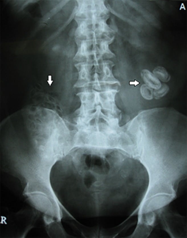
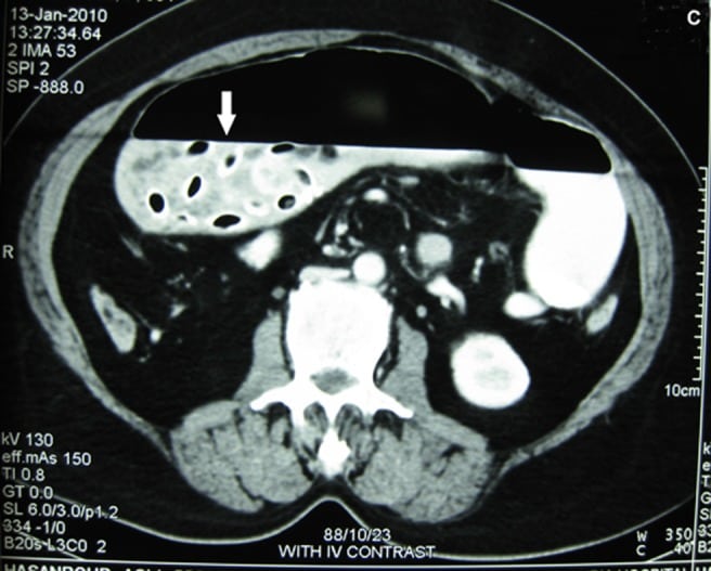
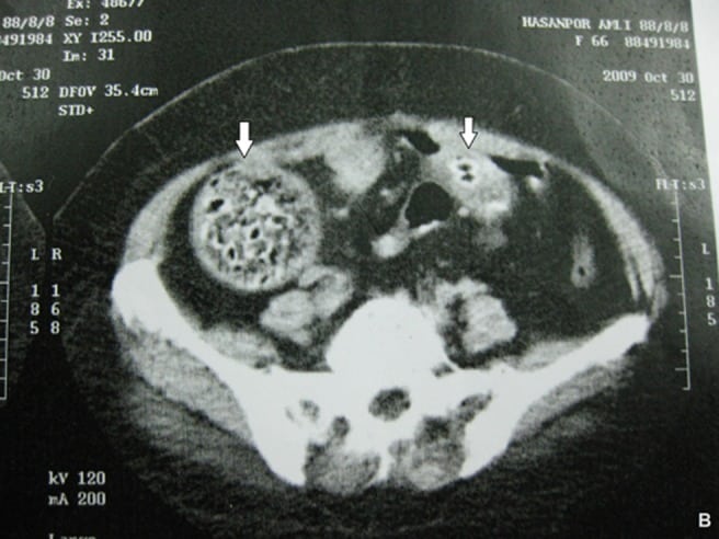
A 55-year-old woman with a 3-month history of abdominal pain presented to the emergency department with chief complaints of worsening abdominal pain and per os intolerance since 3 days ago. Her medical history was noteworthy for watery diarrhea without fever, loss of appetite, weight loss, and nausea and vomiting. Examination revealed abdominal tenderness over right and left lower quadrant (LLQ). Plain radiography and computed tomography showed round calcified mass with small areas of air in cecum and 2.5 and 4.5 cm-diameter round masses in LLQ (Figure 1–3).
Figure 3
Contrast-enhanced abdominal computed tomography transverse image showing round calcified mass with areas of air (arrow).
DISCUSSION
The intestinal obstruction caused by Phytobezoars. Bezoars are classified into 4 main types; phytobezoars, trichobezoars, pharmacobezoars, and lactobezoars. Phytobezoars, the most common types of bezoar, are composed of vegetable matter.1 They are concretion of poorly digested fruit and vegetable fibers that usually become impacted in the narrowest portion of the small bowel.2 Previous gastric resection or ulcer surgery predisposes to bezoar. Abnormal mastication, decreased gastric motility and secretion, autonomic neuropathy in patients with diabetes and myotonic dystrophy are some other predisposing factors.3 The common presenting symptoms are vague feeling of epigastric fullness, nausea, vomiting, anorexia, early satiety, and weight loss.4
The masses were surgically removed in our patient. They consisted of 25–30 foreign bodies in various size including pits, seeds, small pieces of stones, and date kernels.
Figure 2
Contrast-enhanced abdominal computed tomography transverse image showing round calcified mass with areas of air (arrows).
Footnotes
Full text available through open access at http://escholarship.org/uc/uciem_westjem
Address for Correspondence: Seyed Mojtaba Aghili, Imam Khomeini Hospital, Keshavarz Blvd., Vali-asr Sq., Tehran, Iran. Email: m-aghili@tums.ac.ir 7 / 2014; 15:385 – 386
Submission history: Revision received April 11, 2014; Submitted April 23, 2014; Accepted April 28, 2014
Conflicts of Interest: By the WestJEM article submission agreement, all authors are required to disclose all affiliations, funding sources and financial or management relationships that could be perceived as potential sources of bias. The authors disclosed none.
REFERENCES
1 Teng H, Nawawi O, Ng K Phytobezoar: An unusual cause of intestinal obstruction. Biomed Imaging Interv J. 2005; 1:e4
2 Acar T, Tuncal S, Aydin R An unusual cause of gastrointestinal obstruction: Bezoar. N Z Med J. 2003; 116:U422
3 Yildirim T, Yildirim S, Barutcu O Small bowel obstruction due to phytobezoar: Ct diagnosis. Eur Radiol. 2002; 12:2659-2661
4 Mintchev MP Gastric electrical stimulation for the treatment of obesity: From entrainment to bezoars-a functional review. ISRN Gastroenterol. 2013; :434706


