| Author | Affiliation |
|---|---|
| Jiro Tamura, MD | University of the Ryukyus, Department of Infectious, Respiratory, and Digestive Medicine, Okinawa, Japan |
| Nobuo Kuniyoshi, MD | Heart Life Hospital, Surgery Division, Okinawa, Japan |
| Shuichi Maruwaka, MD | Heart Life Hospital, Digestive Division, Okinawa, Japan |
| Joji Shiroma, MD | Heart Life Hospital, Digestive Division, Okinawa, Japan |
| Sunao Miyagi, MD | Heart Life Hospital, Digestive Division, Okinawa, Japan |
| Hitoshi Orita, MD | Heart Life Hospital, Digestive Division, Okinawa, Japan |
| Hiroshi Sakugawa, MD | Heart Life Hospital, Digestive Division, Okinawa, Japan |
| Akira Hokama, MD | University of the Ryukyus, Department of Infectious, Respiratory, and Digestive Medicine, Okinawa, Japan |
| Fukunori Kinjo, MD | University of the Ryukyus Hospital, Department of Endoscopy, Okinawa, Japan |
| Jiro Fujita, MD | University of the Ryukyus, Department of Infectious, Respiratory, and Digestive Medicine, Okinawa, Japan |
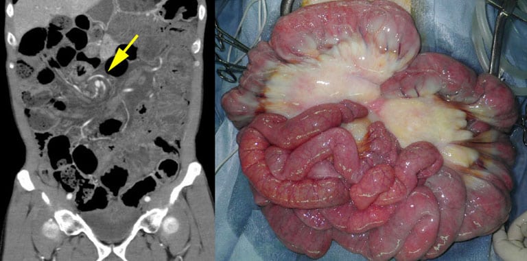
A 59-year-old man had been admitted to our hospital three times with tarry stool, hematemesis, and abdominal discomfort. His medical history included no abdominal operation. Repeated upper endoscopy, colonoscopy, and computed tomography (CT) had been negative. Gastrointestinal bleeding scintigraphy and Meckel scintigraphy had been also negative. In the last admission, he presented abrupt and sharp abdominal pain. An abdominal radiograph showed dilations and air-fluid levels of small intestine and colon. An abdominal CT revealed dilation of small intestine with the lack of contrasts, mesenteric and bowel wall edema, and “clockwise” rotation of the mesentery around the mesenteric vessels (whirl sign) (Figure, arrow). The exploratory laparotomy showed a volvulus of the small intestine at the base of the mesentery, and an edematous mesentery (Figure). The cause of the mesenteric rotation was not identified, such as congenital malrotation, bands, and postoperative adhesion. Primary small bowel volvulus (PSBV) was diagnosed, and the affected bowel was untwisted. Postoperative course was uneventful, and he was discharged home 14 days after surgery.

Figure
Abdominal computed tomography revealed dilation of small intestine with the lack of contrasts, mesenteric and bowel wall edema, and “clockwise” rotation of the mesentery around the mesenteric vessels (whirl sign) (arrow).
PSBV is defined as torsion of large segment small intestine at the basis of the mesentery without any associated underlying cause, such as congenital malrotations, bands, postoperative adhesions, tumors, and diverticular disease. The preoperative diagnosis of PSBV is rather difficult because of limited value of physical examination and radiograph films. However, several authors have reported the usefulness of preoperative abdominal CT for the diagnosis of PSBV.1–3 A tightly twisted mesentery around the point of torsion (whirl sign) was described as a typical sign of volvulus of the small intestine. In conclusion, we emphasize PSBV is an important emergency disease demanding prompt surgical intervention, and whirl sign in CT is the key for its diagnosis.
Footnotes
Full text available through open access at http://escholarship.org/uc/uciem_westjem
Address for Correspondence: Jiro Ramura, MD, University of the Ryukyus, 207 Uehara, Nishihara, Okinawa 903-0215, Japan. Email: jtamura0806@gmail.com. 7 / 2014; 15:359 – 360
Submission history: Revision received January 12, 2014; Submitted January 21, 2014; Accepted April 2, 2014
Conflicts of Interest: By the WestJEM article submission agreement, all authors are required to disclose all affiliations, funding sources and financial or management relationships that could be perceived as potential sources of bias. The authors disclosed none.
REFERENCES
1 Takemura M, Iwamoto K, Goshi S Primary volvulus of the small intestine in an adult, and review of 15 other cases from the Japanese literature. J Gastroenterol. 2000; 35:52-55
2 Kim K, Kim M, Kin S Laparoscopic management of a primary small bowel volvulus: a case report. Surg Laparosc Endosc Percutan Tech. 2007; 17:335-338
3 Birnbaum DJ, Grègoire E, Campan P Primary small bowel volvulus in adult. J Emerg Med. 2013; 44:329-330


