| Author | Affiliation |
|---|---|
| Michael R. Minckler, MD | University of Arizona at South Campus, Department of Emergency Medicine, Tucson, Arizona |
| Cameron Newell, MD | University of Arizona at South Campus, Department of Emergency Medicine, Tucson, Arizona |
| Brian Drummond, MD | University of Arizona at South Campus, Department of Emergency Medicine, Tucson, Arizona |
Patient presentation
Discussion
PATIENT PRESENTATION
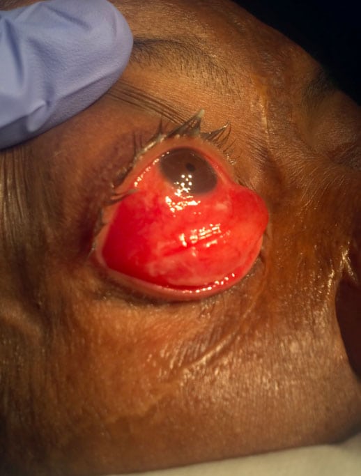
A 51-year-old male with a history of chronic obstructive pulmonary disease and obstructive sleep apnea presents to the emergency department complaining of 48 hours of progressive right eye pain and swelling after he ran into his dresser while sleep-walking. He does not know which surface of the dresser contacted his eye. He denies changes in visual acuity, flashes, or floaters. He has no other complaints and denies recent illness, fever, chills, nausea, vomiting, diarrhea, chest pain, or shortness of breath. Physical exam reveals inability of the patient to close his eyelids on the right. His cranial nerves are intact. His vision exam is unremarkable.
DISCUSSION
The patient has impressive conjunctival chemosis, which is edema of the conjunctiva
This constitutes one half of the reactions that make-up conjunctivitis. It is an inflammatory reaction mediated by the release of histamine, serotonin, and bradykinin. Polymorphonuclear cells also migrate into the reaction site.1
Chemosis is nonspecific finding and is secondary to direct conjunctival endothelial insult. It can result from allergic reaction, bacterial or viral infection, angioedema, or trauma.2 When it is disproportionate to other signs of injury after a traumatic mechanism it can indicate scleral rupture, an ophthalmologic emergency. Chemosis is usually generalized; however, in the setting of scleral rupture it may be confined to only one or two quadrants.3 If there is chronic chemosis following a traumatic mechanism the physician must include lymphatic obstruction most likely secondary to obstructing mass on the differential.4
Globe rupture was not detected in this patient. He could not close his right eyelid, which can lead to ocular damage. A tarsorrhaphy (partially sewing together the eyelids) was performed to protect the eye. At follow-up the patient’s sutures were removed and he was given a pressure patch. By one month this had resolved.
Footnotes
Full text available through open access at http://escholarship.org/uc/uciem_westjem
Address for Correspondence: Michael R. Minckler, MD, University of Arizona at South Campus, Address. Email: Mincklermike1@gmail.com 7 / 2014; 15:357 – 358
Submission history: Revision received February 14, 2014; Accepted March 3, 2014
Conflicts of Interest: By the WestJEM article submission agreement, all authors are required to disclose all affiliations, funding sources and financial or management relationships that could be perceived as potential sources of bias. The authors disclosed none.
REFERENCES
1 Pepperi HF, Ghuman T, Gill KS Conjunctiva. Duane’s Foundations of Clinical Ophthalmology. 2009; chap 29:1
2 Rubenstein JB, Virasch V Allergic conjunctivitis. Ophthalmology. 2008; chap 4.6–4.7
3 Crouch ER, Crouch ER Trauma: ruptures and bleeding. Duane’s Foundations of Clinical Ophthalmology. 2009; chap 61:4
4 Meyer DR Orbital fractures. Duane’s Foundations of Clinical Ophthalmology. 2009; chap 48:2


