
| Author | Affiliation |
|---|---|
| Daniel Weingrow, DO | Arrowhead Regional Medical Center, Departments of Emergency Medicine and Surgery, Colton, California |
| Andrew McCague, DO | Arrowhead Regional Medical Center, Departments of Emergency Medicine and Surgery, Colton, California |
| Ravi Shah, DO | Arrowhead Regional Medical Center, Departments of Emergency Medicine and Surgery, Colton, California |
| Fariborz Lalezarzadeh, DO | Private practice, San Bernardino, California |
ABSTRACT
Volvulus is an unusual condition in Western countries, generally isolated to elderly patients with multiple comorbidities. This report describes an unusual case of a very large gangrenous sigmoid volvulus in a young, otherwise healthy 25-year-old female. A review of the diagnosis and management is subsequently described. Without a consideration of the atypical demographics for sigmoid volvulus, the case illustrates the potential morbidity due to a delayed diagnosis. Early identification and management are crucial in treating sigmoid volvulus before the appearance of gangrene and necrosis, thereby avoiding further complications and associated mortality.
HISTORY
A 25-year-old female, without significant medical history, presented to our hospital emergency department with 5 days of constipation. The patient had previously been admitted to another hospital 2 days before presentation at our institution. At the previous hospital, she received magnesium citrate, sodium phosphate enema, bisacodyl suppository, 4 L of GOLYTELY (Braintree Laboratories Inc, Braintree, Massachusetts), metoclopramide, metronidazole, promethazine, and dicyclomine, along with various analgesics and intravenous fluids. The patient then left the other facility without experiencing improvement. At the time of presentation at our institution, the patient had complaints of diffuse abdominal pain, nausea, vomiting, and persistent constipation but with passage of flatus. The patient reported no fever, dysuria, melena, hematochezia, or recent travel.
On examination, the patient appeared moderately distressed. Breath sounds were clear bilaterally and the patient’s heart was auscultated as a regular rate without murmurs. There was a well-healed anterolateral thoracotomy scar consistent with an unknown cardiac surgery performed shortly after birth. The abdomen was severely distended with diffuse marked tenderness without rigidity or guarding; no hernias or masses were noted and no stool was present in the rectal vault.
The patient’s vital signs were unremarkable. The patient’s laboratory results were notable for a leukocytosis of 18.9 mm3 with a neutrophilic predominance and bandemia of 7%; and sodium levels of 130 mEq/L, potassium levels of 3.3 mEq/L, and bicarbonate levels of 21 mEq/L with an anion gap of 11 mEq/L. Urinalysis was notable for acetonuria and negative β-humanchorionic gonadotropin. Computed tomography (CT) images (Figure 1) were read by the staff radiologist as sigmoid volvulus with severe dilation of the colon.

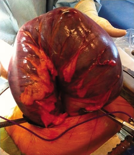
The patient was taken to the operating room for exploratory laparotomy and was found to have 15 to 20 cm of dilated sigmoid colon (Figure 2) with multiple mesenteric twists of sigmoid mesentery with redundant transverse colon. The sigmoid colon was resected with a stapled anastomosis. No intraoperative complications were noted, and the patient was transferred to the surgical intensive care unit. The patient had an uneventful postoperative course and was discharged on postoperative day 7.
DISCUSSION
Volvulus occurs when colon twists on its mesenteric axis with a greater than 180-degree rotation, producing obstruction of intestinal lumen and mesenteric vessels.1 The most common locations for volvulus to occur include the sigmoid colon, cecum, splenic flexure, and transverse colon in order of decreasing frequency.2
Sigmoid volvulus has a variable geographic distribution, being extremely common in developing countries where it affects the young patient, with a lower incidence in Western countries where it predominantly affects the elderly.3 Chronic constipation is blamed for the Western type of sigmoid volvulus, while a high amount of fiber in the diet has been deemed a major factor in the development of sigmoid volvulus in the African population.2,4
Sigmoid volvulus has been classically divided into 2 types, by clinical course, as described by Hinshaw and Carter.5 Acute fulminating volvulus, caused by a complete obstruction, has a clinical presentation of sudden onset periumbilical pain with emesis and constipation. Patients frequently have peritoneal signs on examination.5 Gangrene and perforation are commonly early complications with this type of volvulus. Conversely, with subacute progressive volvulus, patients have only partial obstruction and therefore have a more insidious onset. The subacute form is frequently seen in older patients, with a more subtle clinical picture, described as poorly characterized abdominal cramping, often worse on the left side of the abdomen.5 The understated clinical symptoms in subacute progressive volvulus often lead to delay in diagnosis. On physical examination, upper abdominal distention with associated tenderness, tympany, an empty rectum, and visible peristalsis are all associated with both forms of sigmoid volvulus.
Plain abdominal radiographs may help the diagnosis; however CT, magnetic resonance imaging, and flexible endoscopy are more accurate.6,7 Several radiologic diagnostic signs are described, such as omega or horseshoe sign, bird’s beak sign, Y sign, northern exposure sign, coffee bean sign, bent inner tube or ace of spades sign, left pelvic overlap or left flank overlap sign, liver overlap sign, the whirl sign, and empty left iliac fossa sign.7–9
Surgeons generally advise a 2-step approach, first an endoscopic derotation followed by a subsequent elective surgical correction by colopexy.5,10 Sigmoidosocopy is the initial treatment for those patients without peritoneal signs. Decompression rates vary, with 70% to 90% success. Insertion of a rectal tube should follow to further decompress the viable bowel.3,11,12 Barium enema has been described as another alternative when attempting to untwist a volvulus and is successful in about 5% of patients.13 The disadvantage of sigmoidoscopic decompression includes risk of perforation. Expectant management is not recommended, as spontaneous reduction is found in only 2% of patients and recurrence is high in this group.14
Urgent laparotomy is recommended when decompression is unsuccessful or if the patient is felt to be at high risk for gangrene or perforation. When gangrenous bowel is discovered, immediate resection is necessary.15 After resection, colostomy and mucous fistula, or Hartmann procedure, is recommended.16 In only 10% of cases is colon found to be gangrenous. In cases for which viable colon is encountered, the decision of whether or not to resect must be made. When resected, there is controversy regarding whether to restore intestinal continuity. Generally, if the colon is viable, evidence favors primary anastomosis when feasible.
Nonsurgical detorsion offers the flexibility of scheduling surgery at a next available date.
Some authors suggest a 4-week delay before definitive surgery. Traditional operation is resection of at least the sigmoid colon.3 Laparoscopic resection of the sigmoid colon is growing in popularity and may have a role for high-risk patients or those who may not tolerate conventional surgery.17,18
This report describes sigmoid volvulus in an otherwise healthy young patient with considerable delay in definitive diagnosis. Unfortunately, the patient experienced considerable morbidity because of the unconventional patient profile, traditionally associated with sigmoid volvulus in Western countries. The case emphasizes the importance of early identification in the atypical patient before the appearance of twisted loop gangrene, in order to optimize patient management.
Footnotes
Supervising Section Editor: Sean Henderson, MD
Submission history: Submitted January 31, 2011; Accepted April 11, 2011
Reprints available through open access at http://escholarship.org/uc/uciem_westjem
DOI: 10.5811/westjem.2011.4.6720
Address for Correspondence: Daniel Weingrow, DO
Arrowhead Regional Medical Center, Department of Emergency Medicine, 400 N Pepper Ave, Colton, CA 92324-1819
E-mail: dangrow@gmail.com
Conflicts of Interest: By the WestJEM article submission agreement, all authors are required to disclose all affiliations, funding, sources, and financial or management relationships that could be perceived as potential sources of bias. The authors disclosed none.
REFERENCES
1. Gerwig WH. Volvulus of the colon: symposium on function and disease of anorectum and colon.Surg Clin North Am. 1955. pp. 1395–1399. [PubMed]
2. Northeast ADR, Dennison AR, Lee EG. Sigmoid volvulus: new thoughts on epidemiology. Dis Colon Rectum. 1984;27:260–261. [PubMed]
3. Madiba TE. Thomson SR. The management of sigmoid volvulus. J R Coll Surg Edinb. 2000;45:74–80. [PubMed]
4. Schagen van Leeuen JH. Sigmoid volvulus in a West African population. Dis Colon Rectum.1985;28:712–716. [PubMed]
5. Hinshaw DB, Carter R. Surgical management of acute volvulus of the sigmoid colon: a study of 55 cases. Ann Surg. 1957;146:52–60. [PMC free article] [PubMed]
6. Young WS, Engelbrecht HE, Stocker A. Plain film analysis in sigmoid volvulus. Clin Radiol.1978;29:553–560. [PubMed]
7. Balthazar EJ, Birnbaum BA, Megibow AJ, et al. Closed-loop and strangulating intestinal obstruction: CT signs. Radiology. 1992;185:769–775. [PubMed]
8. Shaff MI, Himmelfarb E, Sacks GA, et al. The whirl sign: a CT finding in volvulus of the large bowel.J Comput Assist Tomogr. 1985;9:410. [PubMed]
9. Hirao K, Kikawada M, Hanyu H, et al. Sigmoid volvulus showing “a whirl sign” on CT. Intern Med.2006;45:331–332. [PubMed]
10. Cirocchi R, Farinella E, La Mura F, et al. The sigmoid volvulus: surgical timing and mortality for different clinical types. World J Emerg Surg. 2010;5:1. [PMC free article] [PubMed]
11. Welch GH, Anderson JR. Acute volvulus of the sigmoid colon. World J Surg. 1987;11:258–262.[PubMed]
12. Bruusgard C. Volvulus of the sigmoid colon and its treatment. Surgery. 1947;22:466–478.[PubMed]
13. Theuer C, Cheadle WG. Volvulus of the colon. Am Surg. 1991;57:145–150. [PubMed]
14. Reilly PMJ, Jones B, Bulkley GB. Volvulus of the colon. In: Cameron JL, editor. Current Surgical Therapy. St Louis, MO: Decker Inc;; 1992. pp. 170–174.
15. Gordon R, Watson K. Ileosigmoid knot. J R Coll Surg Edinb. 1984;29:100–102. [PubMed]
16. Bagarani M, Conde AS, Longo R, et al. Sigmoid volvulus in West Africa: a prospective study in surgical treatments. Dis Colon Rectum. 1993;36:186–190. [PubMed]
17. Pruett B. Laparoscopic colectomy for sigmoid volvulus. J Miss State Med Assoc. 1993. pp. 73–75. [PubMed]
18. Chung CC, Kwok SP, Leung KL, et al. Laparoscopy-assisted sigmoid colectomy for volvulus. Surg Laparosc Endosc. 1997;7:423–425. [PubMed]