
| Author | Affiliation |
|---|---|
| Andrew Little, III, MS | Ohio University College of Osteopathic Medicine, Affinity Medical Center Emergency Department, Massillon, Ohio |
| Andy Culver, DO | Ohio University College of Osteopathic Medicine, Affinity Medical Center Emergency Department, Massillon, Ohio |
ABSTRACT
Diverticulosis is a common disorder among geriatric patients, of whom 10% to 25% go on to develop diverticulitis. Known complications of diverticulitis include formation of phlegmon, fistula, bowel obstruction, bleeding, perforation, and colonic abscess. A less common complication is perforation with formation of an extra-abdominal necrotizing abscess. This case is a report of an 83-year-old female who presented to the emergency department with a necrotizing abdominal wall abscess secondary to right-sided diverticular microperforation.
CASE REPORT
An 83-year-old female presented to the emergency department secondary to an accident involving her wheelchair. Earlier that day she had been seen by her family physician regarding a subcutaneous abscess located on her abdomen and was on her way to see a general surgeon when she had an accident, whereby she was flung from her wheelchair. She was fully immobilized and on a backboard. She was later taken off the backboard after passing Nexus criteria for cervical spine immobilization, with no complaints of pain other than in her left lower quadrant. After being taken off the backboard, a full physical examination was performed. The patient’s vital signs were blood pressure of 124/59, heart rate of 107, respiratory rate of 18, temperature of 97.5°F, and a pulse oximetry of 95% on room air. Auscultation of lungs found them to be clear bilaterally in all fields; cardiac auscultation found normal S1 and S2 with no murmurs, gallops, or rubs. Upon examining the patient’s abdomen, bowel sounds were found in all 4 quadrants and pain was elicited upon palpation of her left lower quadrant, where an 18 × 8-cm subcutaneous abscess was noted. The abscess was fluctuant with a central area of black necrotic skin measuring 8 × 3 cm. Upon direct palpation around the abscess, some oozing was noted centrally. During the physical examination we learned that the patient also suffered from Parkinson disease complicated by dementia, so her husband became our primary historian. Upon questioning him about the mass, he said he had noticed it in the previous 2 days, with the central black necrosis only being present since the previous day. He also stated that his wife had been complaining of increasing pain over the past few days, which was worsened with bowel movements. Upon review of her old medical records, it was noted that she also had a history of gastroesophageal reflux, hypertension, hypothyroidism, renal failure, pulmonary hypertension, diastolic heart failure, and was recently hospitalized for an episode of acute diverticulitis with perforation, for which she was treated medically. With the combination of her presenting symptoms and recent and past medical history, we ordered a complete blood count, basic metabolic panel (BMP), and a urinalysis. An abdominal computed tomography (CT) with intravenous (IV) contrast was ordered. A surgical consult was made while waiting for her laboratory results.
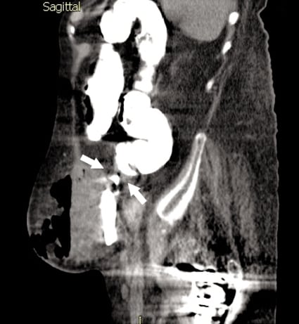
The patient’s laboratory results showed a white blood cell count of 27.9 × 1,000/uL (92% neutrophils, 3.4% lymphocytes, 4.5% monocytes, 0.1% eosinophils, 0.05% basophils), hemoglobin of 9.4 g/dL, hematocrit of 27.6%, and a platelet count of 446. Her BMP showed a serum sodium of 134 mmol/L, potassium of 3.4 mmol/L, blood urea nitrogen of 23 mg/dL, and creatinine of 1.1 mg/dL. Finally, her urinalysis was positive for nitrites, with microscopy showing 1+ leukocytes and 4+ bacteria. The CT revealed an inflamed colon with a fistulous tract winding into the left lower quadrant connecting to a large subcutaneous abscess, with inflammatory changes and pockets of gas and subcutaneous air noted throughout. The patient’s history and her physical examination, laboratory, and CT findings were indicative of necrotizing fasciitis possibly secondary to diverticular perforation (Figure).
A surgical consultant evaluated the patient and agreed with our findings of possible necrotizing infectious process, possibly due to her previous diverticular perforation. The patient was given broad-spectrum antibiotics and IV fluids. Presurgical laboratory tests were performed and she was admitted to the intensive care unit (ICU) and later underwent surgical intervention, which included debridement of large amounts of malodorous pus and fluid consistent with a mixed anaerobic infection. After successful debridement of the abscess, a complete survey of the abscess showed numerous areas of necrosis within the subcutaneous fat, and a small opening at the base of the abscess was noted that emanated pus from the abdominal cavity. This was enlarged for further examination of the patient’s large bowel, where the distal right sigmoid colon was very inflamed and a tiny microperforation was found. Examination of the rest of the large bowel showed no other signs of infection, and the sigmoid microperforation was ruled the source of the patient’s necrotizing infection. The patient then underwent sigmoid resection with placement of a diverting colostomy with wound vacuum placement. Her wound site was cultured during surgery and subsequently grew multiple organisms, which included Proteus mirabilis, Escherichia coli, E faecalis, Bacillus fragilis, and a coagulase-negative staphylococcus. The patient was transferred to the ICU on ventilatory support, where she received medical therapy for sepsis. Her hospitalization was complicated by failure to wean from ventilatory support and by multiple bouts of fever secondary to sepsis. She underwent 3 other surgeries for further abscess debridement. She was later transferred to a long-term acute care hospital for long-term care and rehabilitation.
DISCUSSION
This case is an example of a rare, emergent, important complication of acute diverticulitis.1Typically, patients with acute diverticulitis present with symptoms that include lower left quadrant pain, nausea, vomiting, diarrhea, constipation, flatulence and fever.2 It can also manifest with numerous complications including phlegmon, intra-abdominal abscess, fistulas involving adjacent organs, and distant septicemia.2–4
Necrotizing fasciitis is a condition caused by anaerobic and gram-negative bacteria (ie, Bacteroides,Proteus, and Enterobacter, as in this case). It typically proliferates in areas of trauma, hypoxia, recent surgery, and medical compromise.5 Its presentation is hallmarked by the symptoms of fever, pain out of proportion, crepitance upon palpation of the abscess, and areas of erythema. The diagnoses can be made by incising the suspected abscess; looking for visual clues of necrosis; or by performing imaging to look for free air and/or gas under the skin or in the subcutaneous tissue.5 Care should be taken to recognize necrotizing infections as soon as possible so prompt and proper treatment can be initiated. Owing to the prevalence of diverticulitis, and its high prevalence of perforation in patients older than 60 years, it is very important to get a gastrointestinal history when dealing with anyone presenting with such abdominal complaints.2,3,6 Since necrotizing infections are considered a surgical emergency, it is also important to find the source of infection and consult surgery services emergently so as not to delay surgical intervention.5
As used for this patient, abdominal CT is the best imaging study for ruling in or ruling out colonic involvement.7 When diagnosing acute diverticulitis, CT gives clinicians the most information about location and involvement of adjacent or distant organs or structures when compared to other imaging modalities.3,4 Another imaging modality that could be attempted in this patient would be bedside ultrasonography. Ultrasonography, to the trained user, is a quick, easy, and relatively painless imaging modality that can be used to measure abscess size and to look for channeling abscess contents. Ultrasonography can also be used to differentiate between cellulitis and an actual abscess.8 The limitation of ultrasonography is the difficulty in determining (as in this case) whether or not colonic involvement is present or whether the abscess has penetrated the abdominal wall.3Magnetic resonance imaging (MRI) is a modality that can also be used when investigating intra-abdominal abscesses.9,10 Its effectiveness compared to CT has not been investigated to date, but the utility of MRI is likely comparable to that of CT.11 Some of the issues with using MRI in emergent situations, and other cases of necrotizing infections, are the time necessary to complete the scan (hours instead of minutes) and the availability of scanners.
Treatment for acute diverticulitis is often purely medical, but surgical intervention is indicated in some cases.2,3 Medical management consists of antibiotics targeted to treat common bacteria found in the colon (gram-negative rods and anaerobic bacteria), intravenous fluids, and pain medication while monitoring the patient’s vital signs; management should be started in the emergency department before admission to the hospital. Medical treatment alone is typically reserved for patients who are deemed to be poor surgical candidates and for those who do not suffer from concurrent complications (ie, perforation, bleeding), with a success rate of 70% to 100%.3 Surgical intervention would include treating the patient medically and removing involved portions of the sigmoid colon along with other involved sections of bowel. Patients are typically managed medically first until the complications mentioned above are apparent or are imminent.2,3 The key to treatment of complicated acute diverticulitis is early recognition, early surgical consultation, and initiating treatment in a timely manner.3
Acute diverticulitis is a disease that, as our population ages, is sure to increase in prevalence and presentation to the emergency department. It is important for clinicians to promptly recognize patients who are having an acute episode of diverticulitis so that they can begin treatment and avoid severe complications such as those suffered by the patient in this case study.
Footnotes
Supervising Section Editor: Rick McPheeters, DO
Submission history: Submitted March 23, 2011; Accepted April 8, 2011
Reprints available through open access at http://escholarship.org/uc/uciem_westjem
DOI: 10.5811/westjem.2011.4.6750
Address for Correspondence: Andrew Little, MS
Ohio University College of Osteopathic Medicine, Affinity Medical Center Emergency Department, 875 8th St NE, Massillon, OH 44646
Email: al257807@ohio.edu
Conflicts of Interest: By the WestJEM article submission agreement, all authors are required to disclose all affiliations, funding, sources, and financial or management relationships that could be perceived as potential sources of bias. The authors disclosed none.
REFERENCES
1. Piedra T, Martin-Cuesta L, Arniaz J, et al. Necrotizing fasciitis secondary to diverticulitis. Emerg Radiol. 2007;13:345–348. [PubMed]
2. Nguyen MC, Chudasama YN, Dea SK, et al. Diverticulitis. 2010. eMedicine Web site. Available at:http://emedicine.medscape.com/article/173388-overview. Accessed November 3.
3. Rafferty J, Shellito P, Hyman NH, et al. Practice parameters for sigmoid diverticulitis. Dis Colon Rectum. 2006;49:939–944. [PubMed]
4. Panghaal VS, Chernyak V, Patlas M, et al. CT features of adnexal involvement in patients with diverticulitis. AJR. 2009;192:963–966. [PubMed]
5. Maynor M. Necrotizing fasciitis. 2010. http://emedicine.medscape.com/article/784690-overview eMedicine Web site. Available at. Accessed November 3.
6. Alvarez JA, Baldonedo RF, Bear IG, et al. Presentation, management and outcome of acute sigmoid diverticulitis requiring hospitalization. Dig Surg. 2007;24:471–476. [PubMed]
7. Baker ME. Imaging and interventional techniques in acute left-sided diverticulitis. J Gastrointest Surg. 2008;12:1314–1317. [PubMed]
8. Squire BT, Fox JC, Anderson C. ABCESS: applied bedside sonography for convenient evaluation of superficial soft tissue infections. Acad Emerg Med. 2005;12:601–606. [PubMed]
9. Connolly SA, Connolly LP, Drubach LA, et al. MRI for detection of abscess in acute osteomyelitis of the pelvic in children. AJR. 2007;189:867–872. [PubMed]
10. Balci NC, Semelka RC, Noone TC, et al. Pyogenic hepatic abscesses: MRI findings on T1- and T2- weighted and serial gadolinium-enhanced gradient-echo images. J Magn Reson Imaging.1999;9:285–290. [PubMed]
11. Jung AJ, Yee J, Rosen MP, et al. Palpable abdominal mass: American College of Radiology appropriateness criteria. American College of Radiology. 2011.http://www.acr.org/SecondaryMainMenuCategories/quality_safety/app_criteria/pdf/ExpertPanelongastrointestinalImaging/PalpableAbdominalMassDoc10.aspx Web site. Available at: Accessed March 9.