
| Author | Affiliation |
|---|---|
| Cari E. Matthews, MD | Naval Medical Center, San Diego, CA |
| Vikram K. Garg, MD | Scripps Green Hospital, La Jolla, CA |
| Pallabi Sanyal, MD | Scripps Green Hospital, La Jolla, CA |
| Kandan Baban, DO | Scripps Green Hospital, La Jolla, CA |
| Kenneth Grudko, MD | Scripps Green Hospital, La Jolla, CA |
| Adam G. Field, MD | Naval Medical Center, San Diego, CA |
ABSTRACT
We present an unusual neurologic emergency in an elderly male patient. Given his presentation and risk factors, we presumed the initial symptoms to be secondary to a cerebrovascular accident. As the case evolved, however, it became apparent that a more unusual pathology was present. This case report showcases a rare condition masquerading as a common neurologic emergency.
PATIENT PRESENTATION
We present an unusual neurologic emergency. Our patient was an 85-year-old retired Portuguese fisherman who presented with two days of emesis and one day of diplopia, blurry vision and bilateral ptosis. He also complained of difficulty managing secretions and dysphagia but denied headache, stiff neck, numbness, tingling, extremity weakness or difficulty with bowel or bladder control. Past medical history included emphysema and osteoarthritis. He did not take medications and denied drug allergies. Married for 57 years, he lived with his wife. He had quit using alcohol and tobacco 30 years earlier; prior to that, he had smoked three packs a day. He had no recent sick contacts or travel.
Initial vital signs were as follows: temperature of 98.1°F; blood pressure of 163/75mmHg; pulse of 87 beats per minute; respiratory rate of 16 breaths per minute; oxygen saturation of 98% at room temperature. In general, he appeared ill and dysarthric. His head and neck exam revealed pooling of secretions in the posterior oropharynx, bilateral ptosis. No papilledema was noted. He was oriented to person, place and time. His pupils were equal, round, reactive to light and accommodation. Visual fields were full to confrontation. He had limited abduction of both eyes on testing of extraocular movements. He was limited in his ability to protrude his tongue, although it was midline. Shoulder shrug strength, upper extremity strength and lower extremity strength were 5/5 in the proximal and distal muscle groups. We detected no sensory or proprioceptive deficits. Reflexes were 1+ triceps, biceps, and brachioradialis bilaterally and 2+ patellar and ankle jerks bilaterally. Plantars were downgoing bilaterally. Finger-to-nose testing was intact. Gait was not assessed. The remainder of the physical exam was unremarkable.
Laboratory and imaging studies revealed white cell count of 10.9, hemoglobin of 16.2, hematocrit of 49.1 and platelets of 210. The metabolic panel was unremarkable. Chest radiograph showed a possible early right lower lobe infiltrate. Computed tomography (CT) of the head revealed extensive small vessel disease with old bilateral frontoparietal cerebrovascular events. The magnetic resonance imaging (MRI), including diffusion-weighted imaging, did not reveal any evidence of an acute stroke. Lumbar puncture showed one white cell, zero red cells, 31 protein, and 99 glucose. The gram stain was negative as was the acid-fast bacilli stain. India ink preparation was negative.
In the emergency department (ED), primary consideration was given to patient stability. He was mentating without difficulty, had no respiratory distress, and his airway was patent. As his radiographic studies were inconclusive, we considered additional differential possibilities. Given his age, gender and bulbar symptoms, myasthenia gravis figured prominently. Serological studies for acetylcholine receptor antibodies were sent, but results were not immediately available. Electromyography showed mild slowing of motor and sensory velocities, but these were nonspecific changes. We considered edrophonium testing. This drug acts to inhibit acetylcholinesterase and will improve symptoms in myasthenia gravis by increasing stores of acetylcholine. However, because it may cause marked clinical deterioration and respiratory failure in conditions other than myasthenia, we withheld it, as the patient was stable, and his diagnosis was unclear. He was admitted to the hospital ward for further testing and observation.
Our patient deteriorated precipitously on the first evening of his admission, requiring emergent endotracheal intubation for altered mental status and desaturation. He was completely paralyzed at the time of intubation without medication and was unable to protect his airway, manage secretions, or use the muscles of excursion to facilitate breathing. He remained immobile on the ventilator, despite an early wean of paralytics and sedation. Repeat CT of the head was unchanged.
Five days after his admission, the patient’s 90-year-old wife presented with blurry vision and difficulty swallowing. She was directly admitted to the intensive care unit (ICU) for observation. At that point, we were certain of botulism, given the symptoms in both patients. We contacted the state health department, where our request for release of antitoxin from storage at the Los Angeles International Airport was approved, in cooperation with the Centers for Disease Control (CDC). Both patients received the antitoxin the night the wife was admitted. Despite the antitoxin, the patient’s wife deteriorated clinically and required mechanical intubation. She had two episodes of cardiac arrest during her ICU stay; however, she was able to be resuscitated and weaned from the ventilator.
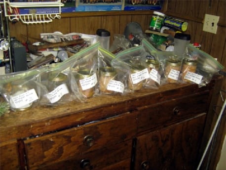
We obtained confirmatory testing late in the ICU course. Blood sent for a botulinum toxin murine bioassay was positive for botulinum toxin A. Stool studies were negative for botulinum toxin. Health department staff went to the patient’s house and collected home-canned tuna jars and home-canned orange juice. The cans were positive for botulinum toxin A.
Approximately two months after discharge from the ICU, the husband was dependent on ventilatory support via tracheostomy and currently resides in a long-term intensive care facility. His wife has regained some strength but has developed dementia and now resides in a skilled-nursing facility.
DISCUSSION
Botulinum toxin is the most potent biologic toxin known. The organism that produces it, Clostridium botulinum, is ubiquitous in the soil. Although several routes of poisoning exist, including wound botulism, which has seen a new emergence in the United States (U.S.) due to intravenous heroin use, and infant botulism via ingestion of spores in a naïve digestive tract, our discussion here focuses on foodborne botulism. Foodborne botulism is caused by the ingestion of preformed toxin that has been expressed by organisms infecting improperly preserved foods. There are seven distinct subtypes, but only A, B, E, and F cause disease in humans. The toxin is inactivated by heating at 85°C for five minutes.1 It is unlikely that our two elderly patients followed these guidelines.
Symptoms begin anywhere from two hours to eight days after ingestion of the toxin, with most patients presenting within 36 hours. Patients present with a classic descending weakness that begins with dysphagia, diplopia and dysarthria. Ptosis, gaze paralysis and facial paralysis are often noted. Motor paralysis progresses from the upper to the lower limbs. In severe poisonings, the diaphragm and intercostals muscles can be affected, causing asphyxiation without ventilator support. Additional findings can include dry mouth, constipation, and dilated pupils. In foodborne botulism, gastrointestinal complaints of nausea and vomiting can occasionally occur prior to neurologic findings. Mental status is unaffected, as are sensory findings. Fever is uncommon.2
ED care is supportive and may include intubation and, in extreme cases, mechanical ventilation. Additional measures include early notification of public health officials to facilitate antitoxin administration and limit ongoing exposures to other potential victims of the contaminated source.
Fewer than 200 cases of all types of botulism are reported in the U.S. annually. Of these, usually 10 to 30 outbreaks occur each year due to improperly processed foods in the home. In the U.S., clinically significant disease is almost universally attributed to subtypes A, B and E. The trivalent antitoxin contains elements to counteract all of these.3 As of March 19, 2010, the CDC has released a heptavalent botulism antitoxin under an investigational drug protocol. It is now the only antitoxin available in the U.S. for naturally occurring non-infant botulism.6
Early administration of antitoxin lends to the most favorable outcomes. Botulinum toxin causes a flaccid paralysis by blocking release of acetylcholine-containing vesicles within the terminal membrane of the motor neuron. This binding at the neuromuscular junction is irreversible; once the condition has progressed to paralysis, improvement will not occur until new motor axons develop to innervate paralyzed muscle fibers.2 Antitoxin will stop disease progression by preventing the serum toxin from binding but will not improve motor function in the affected nerve terminals. Once a patient has become ventilator dependent, it will take months until clinical resolution. Complications such as pneumonia, decubitus ulcers and malnutrition are the rule.
The botulism antitoxin is a horse-serum derived product, so significant allergic reactions are likely to occur; 9% of people may have severe reactions.1 For our patients, we were fortunate to have an allergist available to assist with skin testing prior to antitoxin administration. The antitoxin kit includes instructions and doses for skin testing, so this can be done without the assistance of a specialist, if necessary.
Botulism antitoxin is not routinely stocked in EDs by expert consensus guidelines; it is only accessible via state health departments, whose staffs are able to obtain the antitoxin through the CDC.4 Antitoxin is stocked in strategic prepositioned areas throughout the U.S. The exception to this is the state of Alaska, where the most common botulism poisoning is type E. Trivalent antitoxin is stocked in many municipalities where this poisoning is common, due to native cultural food practices and processing of whale and seal blubbers. Native village health assistants have been trained to recognize the classic pentad of botulism, specifically “nausea and vomiting, dysphagia, diplopia, dry mouth, and dilated and fixed pupils,” and to administer the antitoxin in consultation with state health officials.5
The differential diagnosis for acute weakness in the elderly is broad, and a detailed review is beyond the scope of this article. In this patient population, consideration must be given to etiologies as varied as cerebrovascular accidents (CVA), intracranial mass lesions, dementia, chronic subdural hematomas, depression, adverse drug reactions, thyroid derangements and ingestions. Specific causes of descending paralysis may include myasthenia gravis, pontine cerebrovascular accidents, botulism and Lambert-Eaton syndrome.
These disease processes may be somewhat complicated to distinguish. Myasthenia gravis may be confirmed with symptom improvement after the administration of an acetylcholinesterase inhibitor, such as edrophonium. Brain imaging with CT or MRI would likely be abnormal with a pontine CVA. In Lambert-Eaton syndrome, patients will show an improvement in weakness with repetition of exercise or movement. In fact, the Lambert sign, where grip strength will increase progressively throughout the duration of a hand squeeze, is pathognomic. Botulism can be confirmed with toxin assays; however, because the results will be unavailable in the ED, a careful history must be obtained should this diagnosis be considered.

Footnotes
Supervising Section Editor: Sean Henderson, MD
Submission history: Submitted: April 5, 2010; Revision received June 1, 2010; Accepted June 23, 2010.
Reprints available through open access at http://escholarship.org/uc/uciem_westjem
Address for Correspondence: Cari E. Matthews, MD, Department of Emergency Medicine, Naval Medical Center, San Diego, CA
Email carielaine@yahoo.com
Conflicts of Interest: By the WestJEM article submission agreement, all authors are required to disclose all affiliations, funding sources, and financial or management relationships that could be perceived as potential sources of bias. The authors disclosed none.
REFERENCES
1. Lawrence DT, Dobmeier SG, Bechtel LK, et al. Food poisoning. Emerg Med Clin N Am.2007;25:357–373.
2. Pigott DC. Foodborne illness. Emerg Med Clin N Am. 2008;26:475–497.
3. Kman NE, Nelson RN. Infectious agents of bioterrorism: a review for emergency physicians. Emerg Med Clin N Am. 2008;26:517–547.
4. Dart RC, et al. Expert consensus guidelines for stocking of antidotes in hospitals that provide emergency care. Ann Emerg Med. 2009;54:386–394. [PubMed]
5. Castrodale L. Botulism in Alaska. A guide for physicians and healthcare providers. 2005 Update. State of Alaska. Department of Health and Social Services. Section of Epidemiology.
6. Investigational Heptavalent Botulinum Antitoxin (HBAT) to replace licensed Botulinum antitoxin AB and investigational Botulinum antitoxin E. MMWR. 2010 Mar;59(10):299. [PubMed]