| Author | Affiliation |
|---|---|
| Michael J. Burns, MD | University of California, Irvine School of Medicine, Department of Emergency Medicine, Orange, CA |
| Neel N. Kapadia, MS | University of California, Irvine School of Medicine, Department of Emergency Medicine, Orange, CA |
| Eric F. Silman, MD | University of California, San Francisco, Department of Emergency Medicine |
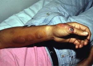
A 25-year-old healthy Hispanic male agricultural laborer presented to the emergency department with six weeks of a painless raised lesion on the proximal thumb with occasional drainage of fluid, without history of injury. Over the next several weeks, he developed painless subcutaneous nodules proximally. He denied any systemic symptoms. The patient emigrated from Mexico two years earlier, but had not traveled since. Physical examination showed an ulcerated, raised, dry, crusted lesion on the lateral surface of the left thumb, with four proximal raised, erythematous, subcutaneous nodules, without epitrochlear or axillary lymphadenopathy (Figure 1). Purulent material was aspirated from one of the nodules; gram, fungal and mycobacterial stains showed no organisms. Saturated solution of potassium iodide (SSKI) was prescribed, and the patient was referred to the Infectious Diseases clinic for follow-up. Thirty days later, the fungal culture grewSporothrix schenkii. The patient was lost to follow-up.

Sporotrichosis is caused by infection with Sporothrix schenkii, a dimorphic fungus, found in soil, wood, and plant surfaces. The fungus is mostly found in the tropics of Central and South America, and Africa.1 The largest U.S. outbreak occurred in 1988, involving 84 people in 15 states, and was associated with exposure to sphagnum moss.2
Lymphocutaneous sporotrichosis, the most common form, presents as a small, nontender, erythematous papulonodule at the site of primary injury. This lesion may be smooth or verrucous, often ulcerates, and develops raised red borders. Over days to weeks, proximal subcutaneous nodules form along the lymphatic drainage, and may ulcerate. Fungal cultures and tissue biopsies aid in the diagnosis.
The differential diagnosis of sporotrichosis includes: nocardiosis, cutaneous leishmaniasis and atypical mycobacterial infection, especially Mycobacterium marinum.3 The treatment of choice for sporotrichosis is oral itraconazole for 3–6 months with SSKI as an alternative. In severe cases, intravenous amphotericin B is used.
Footnotes
Supervising Section Editor: Robert W Derlet, MD
Submission history: Submitted May 07, 2008; Revision Received July 11, 2008; Accepted July 14, 2008
Full text available through open access at http://escholarship.org/uc/uciem_westjem
Address for Correspondence: Michael Burns, MD. Department of Emergency Medicine, 101 The City Dr. Rte 128-01, Orange, CA 92868
Email: mburns@uci.edu
Conflicts of Interest: By the WestJEM article submission agreement, all authors are required to disclose all affiliations, funding sources, and financial or management relationships that could be perceived as potential sources of bias. The authors disclosed none.
REFERENCES
1. Ramos-e-Silva M, Vasconcelos C, Carneiro S, Cestari T. Sporotrichosis. Clin Dermatol.2007;25:181–7. [PubMed]
2. Dixon D, Salkin I, Duncan R, et al. Isolation and characterization of Sporothrix schenckii from clinical and environmental sources associated with the largest U.S. epidemic of sporotrichosis. J Clin Microbiol. 1991;29:1106. [PMC free article] [PubMed]
3. Rex JH, Okhuysen PC. Sporothrix schenckii. In: Mandell GL, Bennett JE, Dolin R, editors. Principles and Practice of Infectious Diseases. 6th ed. Philadelphia, Pennsylvania: Elsevier; 2005. pp. 2984–2988.


