
| Author | Affiliation |
|---|---|
| Azita Javdanfar, PA-C | Keck School of Medicine, University of Southern California, Los Angeles, CA |
| Christopher Celentano, MD | Keck School of Medicine, University of Southern California, Los Angeles, CA |
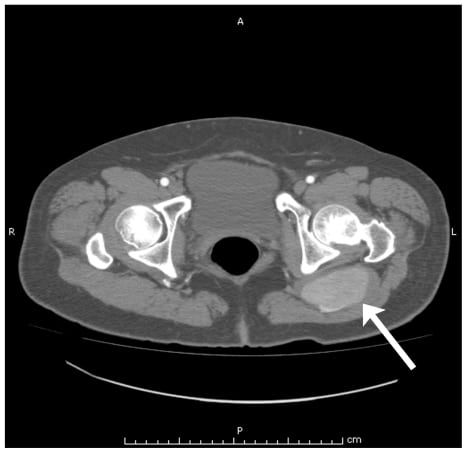
A 59-year-old female with a history of diabetes mellitus presented to the emergency department complaining of three weeks of an enlarging pulsating mass to her left buttock. The patient denied any associated trauma, leg pain, back pain or previous episodes. Physical exam was remarkable for a bounding non-tender pulsatile mass over the lateral left buttock. Skin examination over this area was unremarkable. There were no sensory and motor deficits, and pulses were 2+ distally. A bedside ultrasound of the affected area revealed a pulsatile aneurismal vessel measuring 6cm. Computed tomography (CT) abdomen/pelvis with intravenous contrast confirmed a 7.8 × 4.1 cm aneurysm arising from the left internal iliac artery suggestive of a persistent sciatic artery. A persistent right sciatic artery was also identified on CT of the lower extremities, although there was no evidence of aneurysm noted in that extremity (Figure 1 and and2).2). The vascular surgical service was consulted, and the patient underwent left sciatic aneurysm repair with a polytetrafluoroethylene interposition graft. The patient was discharged from the hospital on post-operative day three without any complications.

Persistent sciatic artery is a rare congenital vascular anomaly estimated to occur in 0.03–0.06% of the population. During embryogenesis, the sciatic artery runs along the sciatic nerve and comprises the major blood supply to the respective lower extremities. As the femoral artery is formed, the bilateral sciatic arteries start to regress. This process continues until the femoral artery completely takes over as the major lower limb blood supply, leaving only remnants of the sciatic artery.1,2 A persistent sciatic artery may occur when there is either a failure of involution of the sciatic artery or failure of the proper development of the femoral artery during normal development. Most cases of persistent sciatic artery are clinically silent and detected only after a vascular complication. Aneurysms develop in approximately a quarter of cases, likely due to the vessel’s anatomic predisposition to minor trauma with sitting. Other complications include thrombosis, embolism and rupture of the artery resulting in ischemia and pain of the lower extremity.2
Ultrasonagraphy and CT are the preferred diagnostic modalities for identifying a suspected sciatic artery aneurysm. CT angiography is useful to delineate the proximal and distal anatomy of the vessel and assist in preoperative planning. A vascular surgeon should be consulted in all cases. Treatment options include surgical exclusion or excision of the aneurysm, endovascular stenting, and endovascular coiling.3
Footnotes
Supervising Section Editor: Eric Snoey, MD
Submission history: Submitted November 4, 2009; Revisions Received: March 22, 2010; Accepted May 10, 2010
Full text available through open access at http://escholarship.org/uc/uciem_westjem
Address for Correspondence: Azita Javdanfar, PA-C, Department of Emergency Medicine, LAC+USC Medical Center, 1200 N State St., Los Angeles, CA 90033
Email: ajavdanfar@yahoo.com
Conflicts of Interest: By the WestJEM article submission agreement, all authors are required to disclose all affiliations, funding sources, and financial or management relationships that could be perceived as potential sources of bias. The authors disclosed none.
REFERENCES
1. Paraskevas G, Papaziogas B, Gigis J, et al. The persistence of the sciatic artery. Folia Morphol.2004;63(4):515–8.
2. Brasileiro JL, Chen J, Santos MA. Persistent sciatic artery aneurysm: case report. J Vasc Bras.2008;7 (1):62–5.
3. Aziz ME, Yusof NRN, Abdullah MS, et al. Bilateral persistent sciatic arteries with unilateral complicating aneurysm. Singapore Med J. 2005;46(8):426–8. [PubMed]