
| Author | Affiliation |
|---|---|
| Warren Johnson, MD | University of Missouri–Kansas City, Children’s Mercy Hospitals and Clinics, Division of Emergency and Urgent Care, Kansas City, Missouri |
| Nirav Shastri, MD | University of Missouri–Kansas City, Children’s Mercy Hospitals and Clinics, Division of Emergency and Urgent Care, Kansas City, Missouri |
| Milton Fowler, MD | University of Missouri–Kansas City, Children’s Mercy Hospitals and Clinics, Division of Emergency and Urgent Care, Kansas City, Missouri |
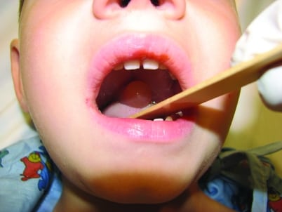
A 4-year-old boy underwent a tonsillectomy and adenoidectomy for tonsillar and adenoidal hypertrophy. The patient developed difficulty breathing after a nonbloody emesis during the car ride home after surgery. The parents noticed a mass in the patient’s mouth and brought him to the clinic. On examination, he had normal vital signs with no respiratory distress and a pulse oximetry of 98% in room air. His oropharynx revealed a markedly edematous, nonerythematous uvula, which was deviated anteriorly and resting on the tongue (see Figure). The tonsillar beds were not bleeding. The rest of his examination yielded normal results.
Isolated angioedema of the uvula is termed Quincke’s disease.1 Causes include trauma, inhalation exposure, for example, marijuana, general anesthesia, medication reaction (angiotensin converter enzyme inhibitors), infections, and hereditary angioedema.1-4
Most reports of uvular edema after ear, nose, and throat procedures are caused by trauma.2 Trauma occurs from the laryngoscope blade during intubation, owing to an oropharyngeal airway, overzealous suctioning, twisting of the uvula during endotracheal tube placement, entrapment of the uvula between the nasal airway and the endotracheal tube, or owing to pressure from a nasogastric tube.
The immediate treatment of uvular edema depends on the degree of airway compromise. Intravenous line should be established and intubation equipment set up at the bedside. Medications used to reduce swelling include epinephrine, diphenhydramine, cimetidine, and methylprednisolone. Patients with suspected noninfectious cause, who do not respond to the above medications, may have a complement deficiency and should also receive plasminogen inhibitor ε-aminocaproic acid.3,4
Our patient received intravenous methylprednisolone, diphenhydramine, and intramuscular epinephrine. He was admitted overnight for observation and discharged home the next day as his symptoms improved significantly. Our patient was not tested for any underlying hereditary cause.
Footnotes
Supervising Section Editor: Sean Henderson, MD
Submission history: Submitted March 15, 2011; Revision received March 16, 2011; Accepted March 21, 2011
Reprints available through open access at http://escholarship.org/uc/uciem_westjem
DOI: 10.5811/westjem.2011.3.2246
Address for Correspondence: Warren Johnson, MD
University of Missouri–Kansas City, Children’s Mercy Hospitals and Clinics, Division of Emergency and Urgent Care, 2401 Gillham Rd, Kansasb City, MO 64108
E-mail: wcjohnson@cmh.edu
Conflicts of Interest: By the WestJEM article submission agreement, all authors are required to disclose all affiliations, funding sources, and financial or management relationships that could be perceived as potential sources of bias. The authors disclosed none.
REFERENCES
1. Deutsch ES, Zalzal GH. Quincke’s edema, revisited. Arch Otolaryngol Head Neck Surg.1991;;117:100–102. [PubMed]
2. Diaz JH. Is uvular edema a complication of endotracheal intubation? Anesth Analg.1993;;76:1139–1141. [PubMed]
3. Goldber R, Lawton R, Newton E, Line WS., Jr Evaluation and management of acute uvular edema.Ann Emerg Med. 1993;;22:251–255. [PubMed]
4. Haselby KA, McNiece WL. Respiratory obstruction from uvular edema in a pediatric patient.Anesth Analg. 1983;;62:1127–1128. [PubMed]