
| Author | Affiliation |
|---|---|
| Akira Hokama, MD | University of the Ryukyus, Department of Infectious, Respiratory, and Digestive Medicine, Nishihara, Okinawa, Japan |
| Shingo Arakaki, MD | University of the Ryukyus, Department of Infectious, Respiratory, and Digestive Medicine, Nishihara, Okinawa, Japan |
| Daisuke Shibata, MD | University of the Ryukyus, Department of Infectious, Respiratory, and Digestive Medicine, Nishihara, Okinawa, Japan |
| Tatsuji Maeshiro, MD | University of the Ryukyus, Department of Infectious, Respiratory, and Digestive Medicine, Nishihara, Okinawa, Japan |
| Fukunori Kinjo, MD | University of the Ryukyus, Department of Endoscopy, Nishihara, Okinawa, Japan |
| Jiro Fujita, MD | University of the Ryukyus, Department of Infectious, Respiratory, and Digestive Medicine, Nishihara, Okinawa, Japan |
ABSTRACT
In emergency, ultrasound has been widely used as a noninvasive and effective examination to evaluate congestive heart failure. We highlight “Playboy Bunny” sign as a reliable marker and an important clue to the diagnosis of passive hepatic congestion, caused by congestive heart failure.
In emergency, ultrasonography has been widely used as a noninvasive and effective examination to evaluate congestive heart failure. The typical ultrasonographic findings in congestive heart failure include hepatomegaly and dilated inferior vena cava. We herein present another impressive sign.
Figure 1 was from a 32-year-old man with congestive heart failure, which was caused by tricuspid regurgitation, mitral valve prolapse, and pericardial effusion. The dilatation of the inferior vena cava and the hepatic veins resembles the “Playboy Bunny” sign. Figure 2 was from a 62-year-old woman with tricuspid regurgitation and pulmonary hypertension who presented with congestive heart failure. The dilated hepatic veins again look like the “Playboy Bunny” sign. Elevated central venous pressure is directly transmitted from the right atrium to the hepatic veins owing to a close anatomic relationship.1 Impaired hepatic venous drainage occurs secondary to cardiac disease, including congestive heart failure, constrictive pericarditis, pericardial effusion, cardiomyopathy, or right-sided valvular disease involving the tricuspid or pulmonary valve. Subsequent dilatation of the inferior vena cava and the hepatic veins then produces the “Playboy Bunny” sign, a good hallmark in passive hepatic congestion.
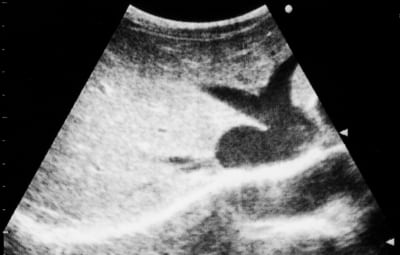
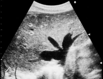
Although Bartrum and Crow2 first described “Playboy Bunny” appearance, with the head being the inferior cava and the ears the hepatic veins, in a normal subject, “Playboy Bunny” sign has been used as an impressive hallmark in passive hepatic congestion. More dilated hepatic veins often present a “deer-horn” appearance.3 In conclusion, we highlight “Playboy Bunny” sign as a reliable marker and an important clue to the diagnosis of passive hepatic congestion, caused by congestive heart failure, in emergency ultrasonography. Although final diagnosis of the type of heart failure is based on clinical and laboratory data, specific ultrasonographic features help narrow the differential diagnosis.
Footnotes
Supervising Section Editor: Sean Henderson, MD
Submission history: Submitted February 14, 2011; Accepted February 28, 2011
Reprints available through open access at http://escholarship.org/uc/uciem_westjem
DOI: 10.5811/westjem.2011.2.2226
Address for Correspondence: Akira Hokama, MD
University of the Ryukyus, Department of Infectious, Respiratory, and Digestive Medicine, 207 Uehara, Nishihara, Okinawa 903-0215, Japan
E-mail: hokama-a@med.u-ryukyu.ac.jp
Conflicts of Interest: By the WestJEM article submission agreement, all authors are required to disclose all affiliations, funding sources, and financial or management relationships that could be perceived as potential sources of bias. The authors disclosed none.
REFERENCES
1. Gore RM, Mathieu DG, White EM, et al. Passive hepatic congestion: cross-sectional imaging features. AJR Am J Roentgenol. 1994;;162:71–75. [PubMed]
2. Bartrum RJ, Jr, Crow HC. Basic ultrasound anatomy. In: Bartrum Jr RJ, Crow HC, editors. Real-Time Ultrasound. 2nd ed. Philadelphia, PA: WB Saunders;; 1983. pp. 74–82.
3. Akdemir R, Yildiz A, Bulur S, et al. Deer horn image in the liver associated with giant right atrium.Am J Geriatr Cardiol. 2007;;16:200–201. [PubMed]