| Author | Affiliation |
|---|---|
| Matthew Silver, MD | Kaiser Permanente, Department of Emergency Medicine, San Diego, California |
| Cyril Thomas, MS PA-C | Kaiser Permanente, Department of Emergency Medicine, San Diego, California |
| Erica Montgomery, PA-S | University of Southern California, Keck School of Medicine, Los Angeles, California |
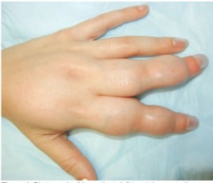
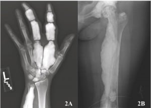
A 38-year-old Hispanic woman with no known past medical or family history presented to the emergency department with severe, intractable left upper and lower extremity pain and inability to walk for 2 days. The woman reported a history of chronic, progressive left hand, arm, and leg deformity over the previous 2 years with episodic flares of severe pain. The woman had not reported any trauma or systemic symptoms. Physical exam revealed deformity and hypertrophy of the second and third digits of the left hand (Figure 1), the left elbow, left thigh, and lower aspect of her left leg without significant joint swelling, warmth or redness. She exhibited significantly limited and painful range of motion at the joints in the left upper and lower extremity. Plain radiographs were obtained (Figure 2).

Photograph of the patients left hand demonstrating marked deformity and apparent swelling of the 2nd and 3rd digits and metacarpal bones.

(A) Radiograph of the left hand showing dense sclerosis, lengthening, and expansion of the second and third phalanges and metacarpals with increased density of the lunate, capitate, and trapezoid and, (B) radiograph of the left proximal femur showing diffuse dense cortical thickening with characteristic “flowing candle wax” appearance.
DIAGNOSIS
Melorheostosis was first described by Leri and Joanny in 1922.1 It is a rare osteosclerotic bone dysplasia caused by a non-inheritable, developmental error in the LEMD3 gene.2 Its incidence is about 0.9 per million and effects males and females equally.3 The pattern of the dysplasia generally follows a sclerotomal distribution, representing the zones of the skeleton supplied by individual spinal sensory nerves, and is typically asymmetric involving the extremities and rarely the axial skeleton. The name melorheostosis is derived from its characteristic “melting wax flowing down a candlestick” appearance on radiologic exam.3
Common presenting symptoms include pain, edema, stiffness, and deformity. The treatment of melorheostosis includes symptomatic treatment for pain, surgery to correct deformities, and amputation if necessary.
Our patient was admitted to the hospital for pain management. Two days later she was discharged to a skilled nursing facility for continued physical therapy, occupational therapy, and medical management.
Footnotes
Supervising Section Editor: Sean O. Henderson, MD
Submission history: Submitted May 24, 2012; Accepted June 25, 2012
Full text available through open access at http://escholarship.org/uc/uciem_westjem
DOI: 10.5811/westjem.2012.6.12680
Address for Correspondence: Matthew A. Silver, MD, Kaiser Permanente, San Diego Medical Center, San Diego, CA
Email: Matthew.a.silver@kp.org
Conflicts of Interest: By the WestJEM article submission agreement, all authors are required to disclose all affiliations, funding sources, and financial or management relationships that could be perceived as potential sources of bias. The authors disclosed none.
REFERENCES
1. Hellemans J, Preobrazhenska O, Willaert A, et al. Loss-of-function mutations in LEMD3 result in osteopoikilosis, Buschke-Ollendorff syndrome and melorheostosis. Nat Genet.2004;36(11):1213–18. [PubMed]
2. Murray RO, McCredie J. Melorheostosis and the sclerotomes: a radiologic correlation.Skeletal Radiol. 1979;4(2):57–71. [PubMed]
3. Suresh S, Muthukumar T, Saifuddin A. Classical and unusual imaging appearances of melorheostosis. Clin Radiol. 2010;65(8):593–600. [PubMed]


