| Author | Affiliation |
|---|---|
| Tyng Yu Chuah, MBBChBAO | Penang Hospital, Department of Emergency Medicine, Georgetown, Malaysia |
| Tze Ping Loh, MBBChBAO | National University Hospital, Department of Laboratory Medicine, Singapore, Singapore |
| Hoi Yin Loi, MBBS | National University Hospital, Department of Diagnostic Imaging, Singapore, Singapore |
| Keat Hwa Lee, MBBS, MS | Penang Hospital, Department of Emergency Medicine, Georgetown, Malaysia |
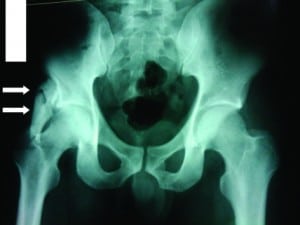
A 35-year-old man presented to the emergency department complaining of right hip pain after being struck by a car while crossing the road. His vital signs were stable, and he complained of right hip pain. He had no other comorbidity. On examination, tenderness and reduced abduction were noted in his right hip, but the gait was normal. The plain radiograph of his pelvis revealed a large, well-circumscribed, ossified mass superior to the greater trochanter and lying parallel to the neck of the right femur (Figure). This patient had a mature post-traumatic myositis ossificans of the right gluteal muscle.

Myositis ossificans is a benign condition characterized by abnormal heterotopic bone formation, typically involving the striated muscle and soft tissue.1 It can present incidentally, as in this patient, or acutely with pain, limitation of joint movement, or complications arising from nerve compression. It is important to recognize plain radiographic features of myositis ossificans because it can be mistaken for a malignant condition. Myositis ossificans shows differing radiographic features in different disease stages. Soft tissue swelling and faint peripheral calcification characterizes the early stage (less than 2 to 4 weeks); later (5 to 24 weeks), well-defined calcification develops, which may be associated with coarser central calcification.1,2 After it has fully matured (more than 6 months), a densely calcified lesion, usually parallel to the long axis of adjacent bone, is visible.1,2Features that suggest malignancy over myositis ossificans include central mineralization, attachment to underlying bone, and increasing size with time (myositis ossificans may shrink as it matures).1,2 Laboratory findings are typically normal; however, erythrocyte sedimentation rate and alkaline phosphatase may be elevated during the acute phase.
Treatment is usually conservative with analgesics and physical therapy, and excision is considered when excessive pain, joint limitation, or nerve compression is present. In this case, the patient was treated with analgesics and was discharged well the following day.
Footnotes
Supervising Section Editor: Sean Henderson, MD
Submission history: Submitted January 16, 2011; Accepted January 24, 2011
Reprints available through open access at http://escholarship.org/uc/uciem_westjem
DOI: 10.5811/westjem.2011.1.2193
Address for Correspondence: Chuah Tyng Yu, MBBChBAO,
Penang Hospital, Department of Emergency Medicine, Jalan Residency, Penang, Malaysia, 10450
E-mail: chuahty@yahoo.com
Conflicts of Interest: By the WestJEM article submission agreement, all authors are required to disclose all affiliations, funding sources, and financial or management relationships that could be perceived as potential sources of bias. The authors disclosed none.
REFERENCES
1. Resnick D, Niwayama G. Soft tissues. In: Resnick D, editor. Diagnosis of Bone and Joint Disorders.Philadelphia, PA: WB Saunders Co; 1995. pp. 4491–4622.
2. Tyler P, Saifuddin A. The imaging of myositis ossificans. Semin Musculoskel Radiol. 2010;;14:201–216.


