Volume 16, Issue 5, September 2015.
Jason D. Heiner, MD
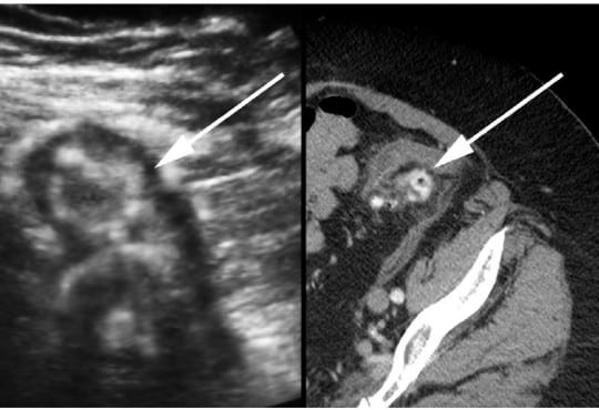
A 72-year-old otherwise healthy female presented to
the emergency department with two weeks of worsening
abdominal pain. She was afebrile with normal vital signs. Her
physical examination was notable for moderate abdominal
tenderness without rebound to the left and suprapubic
regions of the abdomen. Laboratory studies were remarkable
for a white blood cell count of 13,000/mm3
. A focused
bedside ultrasound over the patient’s region of maximal
discomfort revealed a thickened bowel wall and several
small contiguous hypoechoic projections surrounding a
hyperechoic center, suggestive of diverticulitis.
Volume 16, Issue 5, September 2015.
Terri Davis, MSHS, PA-C, et al.
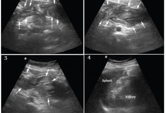
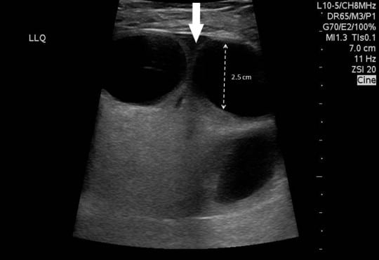
Splenic artery aneurysm rupture is rare and potentially fatal. It has largely been reported in pregnant
patients and typically not diagnosed until laparotomy. This case reports a constellation of clinical and
sonographic findings that may lead clinicians to rapidly diagnose ruptured splenic artery aneurysm
at the bedside. We also propose a rapid, but systematic sonographic approach to patients with
atraumatic hemoperitoneum causing shock. It is yet another demonstration of the utility of bedside
ultrasound in critically ill patients, specifically with undifferentiated shock.
Volume 16, Issue 4, July 2015.
Subramony, MD, et al.
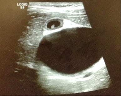
Choledochal cysts are rare but serious bile duct abnormalities are found in young children, usually during the first year of life. They require urgent surgical intervention due to the risk of developing cholangiocarcinoma. Clinicians should consider this diagnosis and perform a point-of-care ultrasound (POCUS) when a child presents to the emergency department (ED) with findings of jaundice, abdominal pain, and the presence of an abdominal mass. We present the case of a six-year-old child presenting only with abdominal pain upon arrival to our ED and was ultimately diagnosed by POCUS to have a choledochal cyst.
Volume 16, Issue 4, July 2015
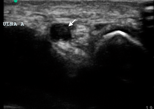
Jonathan Ken, MD, et al.
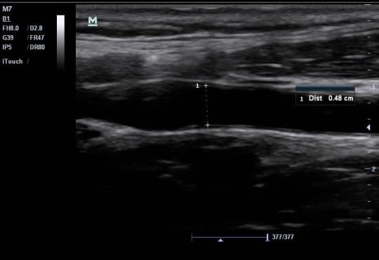
Hypothenar hammer syndrome (HHS) is a rare condition of distal ulnar artery injury and thrombosis secondary to repetitive blunt trauma to the hypothenar area. We present a case of HHS for which point-of-care ultrasound (POCUS) was used as the initial means of imaging, prompting management and disposition without further imaging studies ordered in the emergency department (ED). This case demonstrates the utility of POCUS to aid the Emergency Physician in the diagnosis and management of patients with extremity vascular issues in the ED, and details a rarely seen clinical entity in the ED.
Volume 16, Issue 4, July 2015
Brett Faine, PharmD, et al.
The emergency department (ED) plays a critical role in the management of lifethreatening infection. Prior data suggest that ED vancomycin dosing is frequently inappropriate. The objective is to assess the impact of an electronic medical record (EMR) intervention designed to improve vancomycin dosing accuracy, on vancomycin dosing and clinical outcomes in critically ill ED patients.
Volume 16, Issue 3, May 2015
Jay M. Brenner, MD, et al.
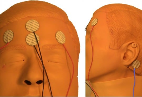
Electroencephalography (EEG) is indicated for diagnosing nonconvulsive status epilepticus (NCSE) in a patient who has altered level of consciousness after a motor seizure. A study in a neonatal population found 94% sensitivity and 78% specificity for detection of seizure using a single-lead device. This study aims to show that a reduced montage EEG would detect 90% of seizures detected on standard EEG.
Volume 16, Issue 3, May 2015
Christopher Gelabert, MD, et al.
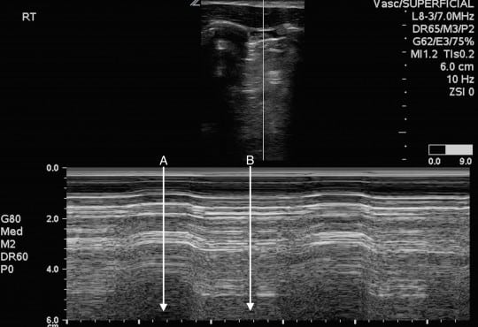
In patients presenting with severe dyspnea, several diagnostic challenges arise in distinguishing the diagnosis of pneumothorax versus several other pulmonary etiologies like bullous lung disease, pneumonia, interstitial lung disease, and acute respiratory distress syndrome. Distinguishing between large pulmonary bullae and pneumothorax is of the utmost importance, as the acute management is very different. While multiple imaging modalities are available, plain radiographs may be inadequate to make the diagnosis and other advanced imaging may be difficult to obtain.
Volume 16, Issue 3, May 2015
Niran Argintaru, MD, et al.
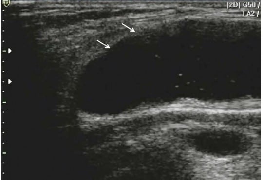
The use of point-of-care ultrasound for the diagnosis of bowel obstructions and hernias is becoming increasingly common in the emergency department (ED). Using a relatively rare case of an incisional port hernia, we demonstrate the ultrasound findings of a strangulated hernia causing a partial small bowel obstruction. A 46-year-old female presented four days following a laparoscopic surgery complaining of abdominal pain, nausea and lack of bowel movements.
Volume 16, Issue 2, March 2015
Cameron Berg, MD et al.
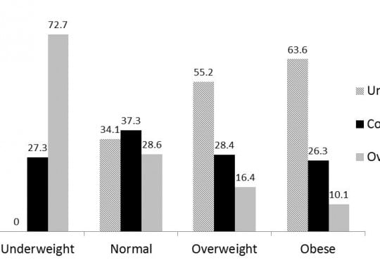
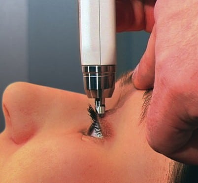
Point-of-care ocular ultrasound (US) is a valuable tool for the evaluation of traumatic ocular injuries. Conventionally, any maneuver that may increase intraocular pressure (IOP) is relatively contraindicated in the setting of globe rupture. Some authors have cautioned against the use of US in these scenarios because of a theoretical concern that an US examination may cause or exacerbate the extrusion of intraocular contents. This study set out to investigate whether ocular US affects IOP. The secondary objective was to validate the intraocular pressure measurements obtained with the Diaton® as compared with standard applanation techniques (the Tono-Pen®).
Volume 16, Issue 2, March 2015
Kevin Padrez, MD et al.
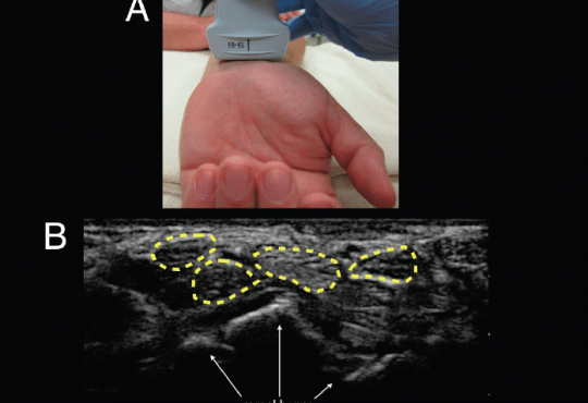
Infectious flexor tenosynovitis (FTS) is a serious infection of the hand and wrist that can lead to necrosis and amputation without prompt diagnosis and surgical debridement. Despite the growing use of point-of-care ultrasound (POCUS) by emergency physicians there is only one reported case of the use of POCUS for the diagnosis of infectious FTS in the emergency department setting. We present a case of a 58 year-old man where POCUS identified tissue necrosis and fluid along the flexor tendon sheath of the hand. Subsequent surgical pathology confirmed the diagnosis of infectious FTS.
Volume 16, Issue 2, March 2015
Lori A. Stolz, MD et al.
Common carotid flow measurements may be clinically useful to determine volume responsiveness. The objective of this study was to assess the ability of emergency physicians (EP) to obtain sonographic images and measurements of the common carotid artery velocity time integral (VTi) for potential use in assessing volume responsiveness in the clinical setting.
Volume 16, Issue 2, March 2015
Srikar Adhikari, MD, MS et al.
Emergency physician-performed compression ultrasonography focuses primarily on the evaluation of the proximal veins of the lower extremity in patients with suspected deep venous thrombosis (DVT). A detailed sonographic evaluation of lower extremity is not performed. The objective of this study was to determine the prevalence of non-thrombotic findings on comprehensive lower extremity venous duplex ultrasound (US) examinations performed on emergency department (ED) patients.
Volume 16, Issue 2, March 2015
Brent Thoma, MD, MA et al.
The number of educational resources created for emergency medicine and critical care (EMCC) that incorporate social media has increased dramatically. With no way to assess their impact or quality, it is challenging for educators to receive scholarly credit and for learners to identify respected resources. The Social Media index (SMi) was developed to help address this.