| Author | Affiliation |
|---|---|
| Francis M. Fesmire, MD | University of Tennessee College of Medicine Chattanooga, Chattanooga, Tennessee |
| Bryan S. England, MD | University of Tennessee College of Medicine Chattanooga, Chattanooga, Tennessee |
| Jared S. Shell, MD | University of Tennessee College of Medicine Chattanooga, Chattanooga, Tennessee |
| Thomas G. Devlin, MD | University of Tennessee College of Medicine Chattanooga, Chattanooga, Tennessee |
| Ron C. Buchheit, MD | University of Tennessee College of Medicine Chattanooga, Chattanooga, Tennessee |
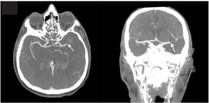
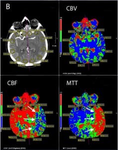
A 43-year-old Caucasian male with history of mechanical mitral valve was found down outside a recreational park shortly after daybreak. Examination on arrival to the emergency department revealed altered mental status, right hemiplegia, forced leftward gaze, and complete aphasia. Patient was ineligible for tissue plasminogen (TPA) therapy due to unknown time of symptom onset. Computed tomography angiogram (CTA) revealed occlusion of the left middle cerebral artery (MCA) with acute thrombus (Figure 1). Computed tomography perfusion scan (CTP) revealed a large ischemic penumbra with no evidence of infarcted brain tissue (Figure 2). The patient was taken for emergent endovascular therapy with successful retrieval of left MCA thrombus. The patient had almost complete resolution of symptoms with a pre-discharge National Institute of Health Stroke Scale of 1. History prior to discharge revealed that the patient was non-compliant and had been off of his warfarin therapy for months prior to the stroke. The patient was discharged home on warfarin and statin therapy.
Figure 1. Transverse (left) and coronal (right) computed tomography angiogram demonstrating abrupt cutoff of the left middle cerebral artery at the site of the thrombus (marked by arrows).
Figure 2. Computer-generated perfusion map demonstrating the region of the left cerebral hemisphere (CBF) with decreased cerebral blood flow and prolonged mean transit time (MTT) (marked by arrows). Note that this region of the brain has normal cerebral blood volume (CBV) indicating potentially salvageable brain tissue.
Acute stroke patients with unknown time of symptom onset are traditionally excluded from acute interventional therapy due to increased rates of fatal intracranial hemorrhage when patients with infarcted brain are treated with TPA or endovascular therapy beyond the recommended time windows.1,2 CTP of the brain is a rapid means of distinguishing between viable and non-viable brain tissue.3 Early in the course of stroke, there is prolonged mean transit time (MTT), decreased cerebral blood flow (CBF), and equal or greater cerebral blood volume (CBV) in ischemic areas of the brain. As brain tissue infarcts, both the CBF and CBV decrease. Assessment of differences between CBF and CBV in areas of brain with prolonged MTT allows one to determine both the size of the stroke as well as the amount of salvageable brain tissue. CTP has the potential to guide acute stroke interventional therapy in patients with unknown time of symptom onset.
Footnotes
Address for Correspondence: Francis Fesmire, MD, University of Tennessee College of Medicine Chattanooga, 960 East Third Street, Suite 100, Chattanooga, TN 37403. Email: francis.fesmire@gmail.com. 2 / 2014; 15:109 – 110
Submission history: Revision received September 9, 2013; Submitted September 16, 2013; Accepted September 16, 2013
Conflicts of Interest: By the WestJEM article submission agreement, all authors are required to disclose all affiliations, funding sources and financial or management relationships that could be perceived as potential sources of bias. Thomas G. Devlin has research funding from Covidian and Brainsgate, speaker’s honoraria from Genetech, and consultant for Codman. The other authors disclosed none.
REFERENCES
1. Stemer A, Lyden P. Evolution of thrombolytic treatment window for acute ischemic stroke. Curr Neurol Neurosci Rep. 2010;10:29–33. [PMC free article] [PubMed]
2. Gurnwald IQ, Wakhloo AK, Walter SM, et al. Endovascular Stoke Treatment Today. Am J Neuroradiol. 2011;32:238–243. [PubMed]
3. Murphy BD, Fox AJ, Lee DH, et al. Identification of penumbra and infarct in acute ischemic stroke using computed tomography perfusion-derived blood flow and volume measurements. Stroke. 2006;37:1771–1776. [PubMed]


