
| Author | Affiliation |
|---|---|
| Shane Davis, MD | Virginia Commonwealth University Health Systems, Department of Emergency Medicine, Richmond, Virginia |
| Angela Creditt, DO | Virginia Commonwealth University Health Systems, Department of Emergency Medicine, Richmond, Virginia |
ABSTRACT
Case Presentation
A 58-year-old female with history of hepatitis C virus presented to the emergency department with a bilateral skin eruption to her feet for one year. Following skin biopsy, the patient was diagnosed with Necrolytic acral erythema (NAE). She was treated with clobetasol ointment, zinc supplementation, and mupirocin, which resulted in improvement in her symptoms.
Discussion
NAE is a rash described as sharply demarcated, lichenified plaques on the dorsal foot and is a rare extra-hepatic manifestation of hepatitis C. This case details a patient with a skin eruption consistent with NAE.
CASE PRESENTATION
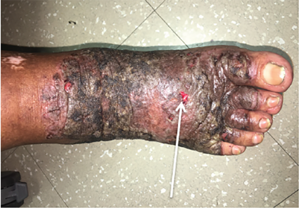
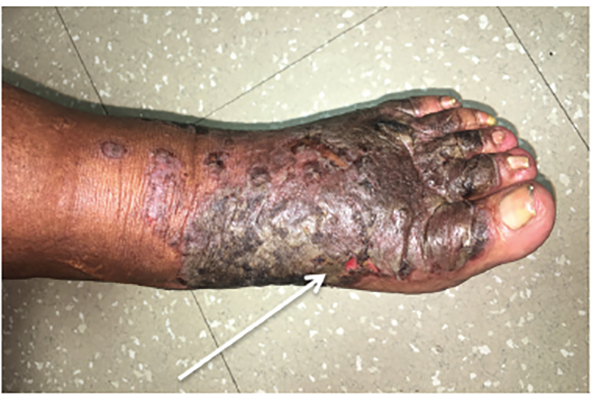
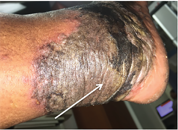
A 58-year-old female with history of hepatitis C virus (HCV) presented to the emergency department with a bilateral skin eruption to her feet for one year. She described it as intermittent and severely painful causing her difficulty with ambulation. Physical exam revealed sharply demarcated, hyperpigmented, lichenified plaques on the dorsa of the feet extending circumferentially around her ankles, as seen in Images 1–3, with areas of fissuring and purulent drainage consistent with superinfection. The patient was started on clindamycin and referred to dermatology. Following skin biopsy, the patient was diagnosed with NAE. She was treated with clobetasol ointment, zinc supplementation, and mupirocin, which resulted in improvement in her symptoms. However, symptoms returned once medications were stopped. The patient was restarted on zinc and clobetasol and referred to hepatology for treatment of her HCV.
DISCUSSION
Necrolytic acral erythema (NAE) is a cutaneous manifestation of hepatitis C virus (HCV) with a prevalence of 1.7% among this patient population.1 NAE is a well-defined, erythematous, tender plaque that typically appears on the dorsa of the feet but can also spread to the posterior ankle. With progression of disease, the appearance becomes thickened and velvety with a surrounding rim of erythema and fissures within the plaque.2 The pathophysiology of NAE remains unclear and the low prevalence rate makes it difficult to determine risk factors. There is speculation that serum or skin zinc deficiency may play a role in this cutaneous manifestation in HCV patients;2 however, there is variable responsiveness of NAE to zinc supplementation, and the only definitive treatment thus far is hepatitis C antivirals.3
CPC-EM Capsule
What do we already know about this clinical entity?
This rash that is an extra-hepatic manifestation of hepatitis C, called necrolytic acral erythema. Although the association is known, the pathophysiology behind the rash is incompletely understood.
What is the major impact of the image(s)?
This rare but characteristic rash that is an extra-hepatic manifestation of hepatitis C virus.
How might this improve emergency medicine practice?
This patient was initially mismanaged due to the rarity of this diagnosis. These images in the emergency medicine literature may potentially aid in proper diagnosis and treatment.
Footnotes
Section Editor: Austin Smith, MD
Full text available through open access at http://escholarship.org/uc/uciem_cpcem
The authors attest that their institution requires neither Institutional Review Board approval, nor patient consent for publication of this image in emergency medicine. Documentation on file.
Address for Correspondence: Angela Creditt, DO, Virginia Commonwealth University Health Systems, Department of Emergency Medicine, 401 N 12th Street, Richmond, VA 23298. Email: angela.creditt@vcuhealth.org. 4:491 – 492
Submission history: Revision received January 9, 2020; Submitted June 27, 2020; Accepted July 3, 2020
Conflicts of Interest: By the CPC-EM article submission agreement, all authors are required to disclose all affiliations, funding sources and financial or management relationships that could be perceived as potential sources of bias. The authors disclosed none.
REFERENCES
1. Raphael BA, Dorey-Stein ZL, Lott J, et al. Low prevalence of necrolytic acral erythema in patients with chronic hepatitis C virus infection. J Am Acad Dermatol. 2012;67(5):962-8.
2. Abdallah MA, Hull C, Horn TD. Necrolytic acral erythema: a patient from the United States successfully treated with oral zinc. Arch Dermatol. 2005;141(1):85-7.
3. Fernandes Botelho LF, Silva Enokihara EMM, Enokihara MY. Necrolytic acral erythema: a rare skin disease associated with hepatitis C virus infection. Anais Brasileiros de Dermatologia. 2016;91(5):649-51.