| Author | Affiliation |
|---|---|
| Thomas M. Nappe, DO | University of South Florida Morsani College of Medicine, Tampa, Florida. Lehigh Valley Hospital, Department of Emergency Medicine, Allentown, Pennsylvania |
| Leonel Diaz, DO | University of South Florida Morsani College of Medicine, Tampa, Florida. Lehigh Valley Hospital, Department of Emergency Medicine, Allentown, Pennsylvania |
| Elizabeth M. Evans, DO | University of South Florida Morsani College of Medicine, Tampa, Florida. Lehigh Valley Hospital, Department of Emergency Medicine, Allentown, Pennsylvania |
CASE
A 56-year-old female presented to the emergency department (ED) with a chief complaint of urinary retention and overflow incontinence for 24 hours, preceded by progressive difficulty with voiding, worsening lower abdominal discomfort and bloating. Her past medical history was significant for small bowel obstruction and neurofibromatosis with an associated benign pelvic tumor that caused similar symptoms as a child, but had been known to be stable since that time. She had also recently been treated for a urinary tract infection. Her physical exam revealed tachycardia and a diffusely tender abdomen with a palpable, tender suprapubic mass extending just above her umbilicus.
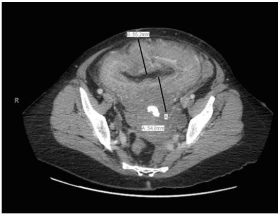
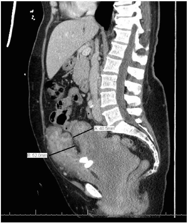
Bedside ultrasonography was performed to visualize her kidneys and bladder and revealed bilateral hydronephrosis and a distended bladder with marked wall thickening (video), yielding further evaluation with computed tomography (CT). A urinary catheter was placed and 1,850 milliliters of urine were collected. CT of the abdomen and pelvis then confirmed bilateral hydronephrosis and a severely enlarged bladder with a diffusely thickened wall, consistent with the nodular appearance expected with neurofibroma of the bladder, as demonstrated in the figure. Laboratory analysis of urine and blood supported suspicion of urinary tract infection and obstructive uropathy, respectively. Histopathological analysis subsequently confirmed the presence of a neurofibroma.
DISCUSSION
Neurofibroma of the bladder is an extremely rare manifestation of neurofibromatosis type 1, or von Recklinghausen disease, not typically seen in the ED.1,2 Although the bladder is the most commonly affected site within the genitourinary system, there are less than 80 reported cases in the literature.2 As in our patient, neurofibroma of the bladder can lead to obstructive uropathy with hydronephrosis.3 Here, we used bedside ultrasonography in the ED for evaluation of symptomatology, which led to the preliminary diagnosis of this rare manifestation, further captured by CT.
Footnotes
Section Editor: Sean O. Henderson, MD
Full text available through open access at http://escholarship.org/uc/uciem_westjem
Address for Correspondence: Thomas M. Nappe, DO, Lehigh Valley Hospital-Muhlenberg, 5th Floor, South Wing, Bethlehem, PA 18017. Email: Tom.nappe@gmail.com. 9 / 2015; 16:756 – 757
Submission history: Revision received May 26, 2015; Accepted June 15, 2015
Conflicts of Interest: By the WestJEM article submission agreement, all authors are required to disclose all affiliations, funding sources and financial or management relationships that could be perceived as potential sources of bias. The authors disclosed none.
REFERENCES
1. Cheng L, Scheithauer BW, Leibovich BC, et al. Neurofibroma of the urinary bladder. Cancer. 1999;86(3):505-13.
2. Üre I, Gürocak S, Gönül II, et al. Neurofibromatosis Type 1 with Bladder Involvement. Case Rep Urol. 2013;2013:145076.
3. Wong-You–Cheong JJ, Woodward PJ, Manning MA, et al. Neoplasms of the Urinary Bladder: Radiologic-Pathologic Correlation. Radio Graphics. 2006;26(2):553-80.
4. Kanyilmaz S, Calis FA, Cinar Y, et al. Bladder wall thickness and ultrasound estimated bladder weight in healthy adults with portative ultrasound device. J Res Med Sci. 2013;18(2):103-6.


