
| Author | Affiliation |
|---|---|
| Shadi Lahham, MD, MS | University of California, Irvine, Department of Emergency Medicine, Irvine, California |
| Samer Assaf, MD | University of California, Irvine, Department of Emergency Medicine, Irvine, California |
| Romeo Fairley, MD | University of California, Irvine, Department of Emergency Medicine, Irvine, California |
CASE REPORT
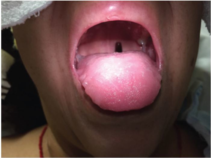
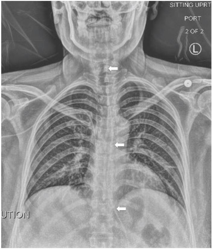
A 41-year-old female presented to the emergency department with nausea, vomiting and foreign body sensation in her throat. The patient had multiple co-morbidities including hypertension, diabetes, cervical cancer and gastroparesis with gastrojejunostomy (GJ) tube. The patient had stable vitals, was in no respiratory distress, and her only complaint was mild throat pain and abdominal pain at the GJ tube insertion site. Physical exam revealed a foreign object in the oropharynx (Figure 1). Abdominal exam showed a soft, non-distended, non-tender abdomen with GJ-tube and colostomy in place. Abdominal series and upright chest radiograph were obtained (Figure 2).
DIAGNOSIS
Mal-positioned GJ tube. Oral exam showed the distal end of the GJ tube protruding into the oropharynx (Figure 1). Upright chest radiograph showed the GJ tube extending superiorly up the esophagus into the oropharynx (Figure 2).
A GJ tube is a percutaneous device that provides access to both the stomach and jejunum1,2. This tube is positioned at the same location as a gastric feeding tube but is longer in order to reach the jejunum. Its purpose is to provide decompression of the stomach and enteric feeding to patients with poor caloric intake.3 The rate of complications of GJ tubes vary between 1–13%.4,5 Many of these complications are considered minor with <1% causing mortality.6-9 In patients with vomiting there is a chance that the GJ tube is displaced from the jejunum and can enter into the esophagus. This can be confirmed with chest radiograph or CT chest.10,11
Footnotes
Section Editor: Sean O. Henderson, MD
Full text available through open access at http://escholarship.org/uc/uciem_westjem
Address for Correspondence: Shadi Lahham, MD, MS, University of California, Irvine, Department of Emergency Medicine, 333 The City Boulevard West, Suite 640, Rt 128-01, Orange, CA 92868. Email: slahham@uci.edu. 12 / 2015; 16:1199 – 1200
Submission history: Revision received September 1, 2015; Accepted September 3, 2015
Conflicts of Interest: By the WestJEM article submission agreement, all authors are required to disclose all affiliations, funding sources and financial or management relationships that could be perceived as potential sources of bias. The authors disclosed none.
REFERENCES
1. Pearce CB, Duncan HD. Enteral feeding. Nasogastric, nasojejunal, percutaneous endoscopic gastrostomy, or jejunostomy: its indications and limitations. Postgrad Med J. 2002;78:198-204.
2. Stroud M, Duncan H, Nightingale J. Guidelines for enteral feeding in adult hospital patients. Gut. 2003;52(Suppl 7):vii1-vii12.
3. Malmgren A, Hede GW, Karlström B, et al. Indications for percutaneous endoscopic gastrostomy and survival in old adults. Food Nutr Res. 2011;55.
4. Larson DE, Burton DD, Schroeder KW, et al. Percutaneous endoscopic gastrostomy. Indications, success, complications and mortality in 314 consecutive patients. Gastroenterology. 1987;93:48-52.
5. Lynch CR, Fang JC. Prevention and management of complications of percutaneous endoscopic gastrostomy tubes. Practical Gastroenterol. 2004;22:66-76.
6. DeLegge RL, DeLegge MH. Percutaneous endoscopic gastrostomy evaluation of device materials: are we “failsafe”?. Nutr Clin Pract. 2005;20:613-617.
7. Milanchi S, Wilson MT. Malposition of percutaneous endoscopic-guided gastrostomy: Guideline and management. J Minim Access Surg. 2008;4:1-4.
8. Sridhar AV, Nichani S, Luyt D, et al. Candida peritonitis: A rare complication following early dislodgement of percutaneous endoscopic gastrostomy tube. J Pediatr Child Health. 2006;42:145-6.
9. Nataraja RM, Fishman JR, Naseer A, et al. Percutaneous endoscopic gastrostomy placement in a human immunodeficiency virus-positive pediatric population leads to an increase in minor complications. J Laparoendosc Adv Surg Tech A. 2011;21:171-175.
10. Timratana P, El-Hayek K, Shimizu H, et al. Percutaneous endoscopic gastrostomy (PEG) with T-fasteners obviates the need for emergent replacement after early tube dislodgement. Surg Endosc. 2012;26:3541-3547.
11. Schrag SP, Sharma R, Jaik NP, et al. Complications related to percutaneous endoscopic gastrostomy (PEG) tubes. A comprehensive clinical review. J Gastrointestin Liver Dis. 2007;16:407-418.