
| Author | Affiliation |
|---|---|
| Faraz Khan, MS | University of California, Irvine School of Medicine, Irvine, California |
| Shadi Lahham, MD, MS | University of California, Irvine, Department of Emergency Medicine, Orange, California |
CASE PRESENTATION
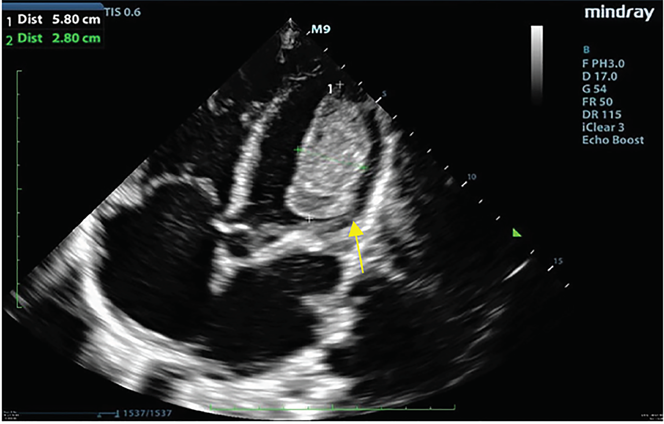
A 34-year-old female with a history of methamphetamine-associated cardiomyopathy presented to the emergency department (ED) with generalized weakness, altered mental status, and chest pain. She reported a recent placement of an automatic implantable cardioverter-defibrillator at an outside hospital three months prior to current presentation and had a documented ejection fraction of 15%. Upon arrival to the ED, she was hypotensive with a systolic blood pressure ranging in the 40s to 70s millimeters of mercury and was hypothermic at 33.6 degrees Celsius. She appeared cachectic and had a 3/6 systolic ejection murmur at the left upper sternal border. We performed a point-of-care ultrasound (POCUS) to assess the patient’s cardiac function and found a large left ventricular (LV) thrombus measuring 5.8 × 2.8 centimeters (Image). Further views of the thrombus seen in the video reveal a large hyperechoic density in the left ventricle. The patient was admitted to the intensive care unit for vasopressor support and thrombolytic therapy.
DISCUSSION
Cardiovascular disease is the leading cause of death in patients with methamphetamine use, and cardiomyopathy is a rare complication that can occur.1 This can lead to systolic dysfunction and reduced ejection fraction, which is an important risk factor for the formation of LV thrombi.2 In patients with methamphetamine-associated cardiomyopathy with an ejection fraction less than 40%, up to 33% can develop a LV thrombus.3 POCUS can be used to help diagnose patients with an LV thrombus.4 Patients found to have a thrombus should be started on anticoagulation therapy.5
CPC-EM Capsule
What do we already know about this clinical entity?
Left ventricular thrombus is a complication of cardiomyopathy and can present with shortness of breath and fatigue.
What is the major impact of the image(s)?
These images depict a left ventricular thrombus as seen on point-of-care ultrasound (POCUS).
How might this improve emergency medicine practice?
Emergency physicians can use POCUS to quickly identify a left ventricular thrombus.
Footnotes
Section Editor: Christopher Sampson, MD
Full text available through open access at http://escholarship.org/uc/uciem_cpcem
Documented patient informed consent and/or Institutional Review Board approval has been obtained and filed for publication of this case report.
Address for Correspondence: Shadi Lahham, MD, MS, University of California, Irvine, Department of Emergency Medicine, 333 City Blvd. West, Suite 640, Orange, CA 92868. Email: slahham@uci.edu. 3:65 – 66
Submission history: Revision received August 29, 2018; Submitted November 26, 2018; Accepted January 1, 2019
Conflicts of Interest: By the CPC-EM article submission agreement, all authors are required to disclose all affiliations, funding sources and financial or management relationships that could be perceived as potential sources of bias. The authors disclosed none.
REFERENCES
1. Paulus M. Methamphetamine use disorder: Epidemiology, clinical manifestations, course, assessment, and diagnosis. UpToDate. Available at: https://www.uptodate.com/contents/methamphetamine-use-disorder-epidemiology-clinical-manifestations-course-assessment-and-diagnosis. Accessed July 30, 2018.
2. Kalaria VG, Passannante MR, Shah T. Effect of mitral regurgitation on left ventricular thrombus formation in dilated cardiomyopathy. Am Heart J. 1998;135(2 Pt 1):215-20.
3. Schürer S, Klingel K, Sandri M, et al. Clinical characteristics, histopathological features, and clinical outcome of methamphetamine-associated cardiomyopathy. JACC Heart Fail. 2017;5(6):435-45.
4. Ünlüer EE, Payza U, Bayata S, et al. Left ventricular thrombus diagnosed by point-of-care ultrasonography. J Emerg Trauma Shock. 2016;9(1):42-3.
5. Colucci WS, Lip GYH. Antithrombotic therapy in patients with heart failure. UpToDate. Available at: https://www.uptodate.com/contents/antithrombotic-therapy-in-patients-with-heart-failure. Accessed July 30, 2018.
SUPPLEMENTARY MATERIAL
A hyperechoic mobile structure within the left ventricle is seen using point-of-care ultrasound through the parasternal long axis, parasternal short axis, and apical four-chamber views of the heart.