
| Author | Affiliation |
|---|---|
| Melissa Myers, MD | San Antonio Military Medical Center, Department of Emergency Medicine, San Antonio, Texas |
| Jared Cohen, MD | San Antonio Military Medical Center, Department of Emergency Medicine, San Antonio, Texas |
CASE PRESENTATION
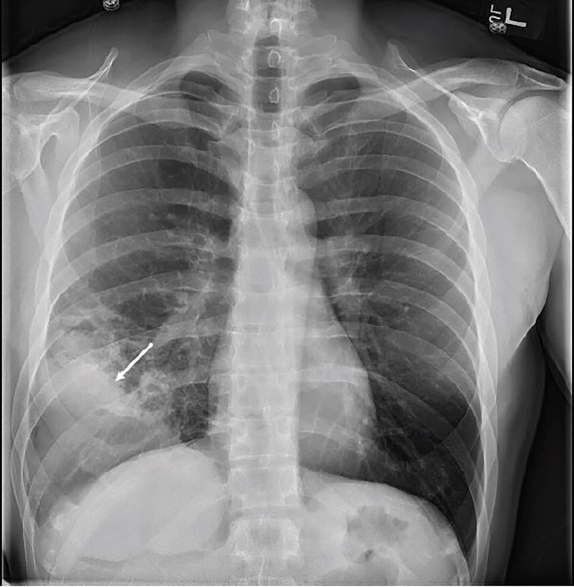
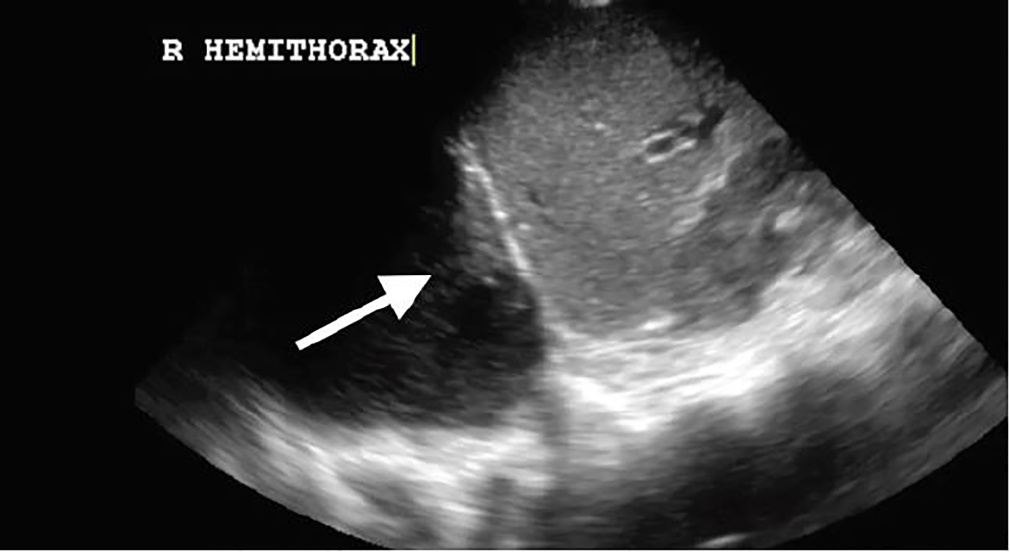
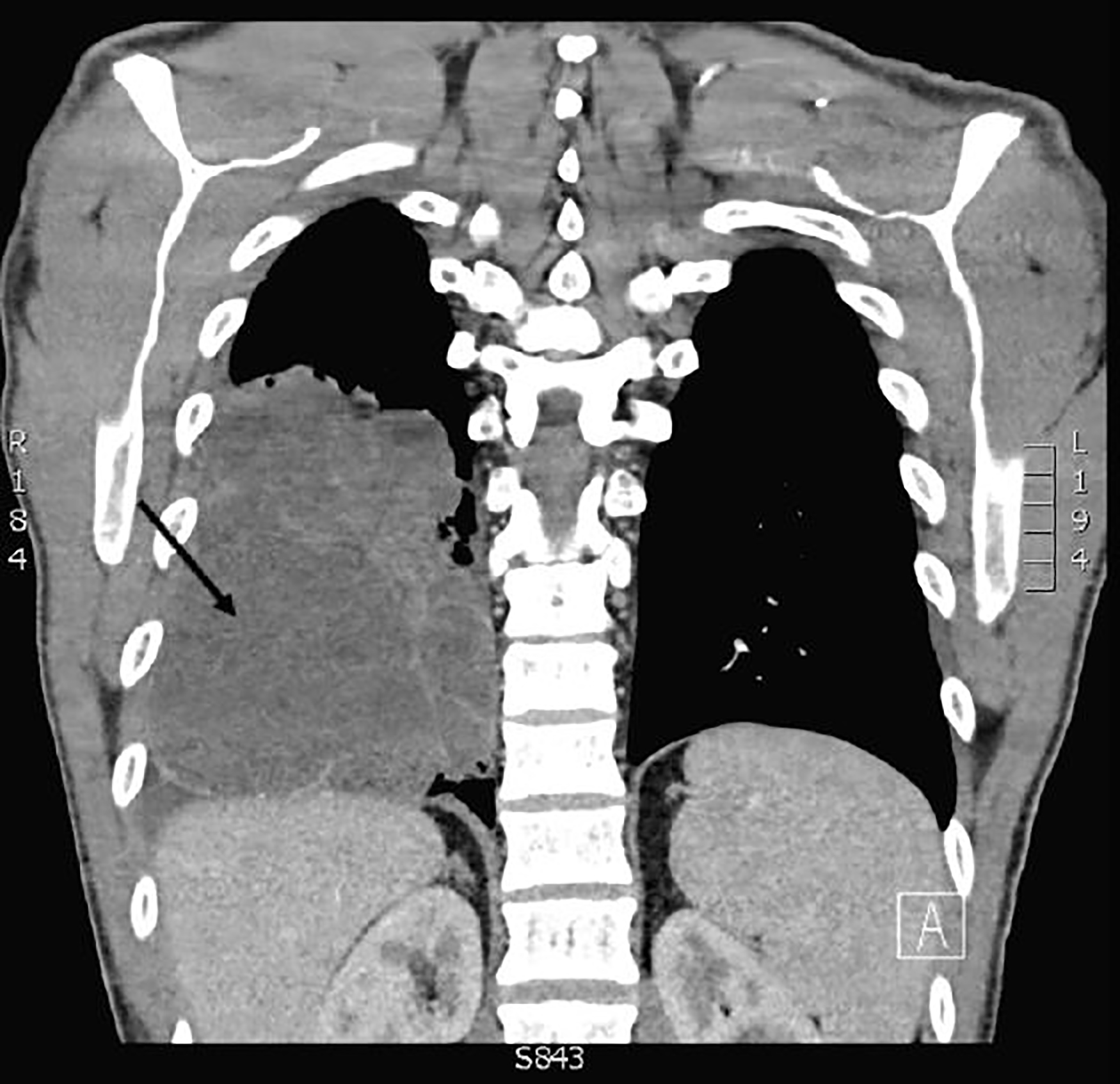
A 35-year-old, otherwise-healthy male presented to a military emergency department complaining of persistent cough. He had been treated for community-acquired pneumonia three weeks prior to this presentation with a five-day course of azithromycin. His vital signs were stable with no fever, hypoxemia or respiratory distress. His physical exam was unremarkable with no focal lung findings. Chest radiograph (Image 1) was performed and was concerning for consolidation or effusion. Pulmonary point-of-care ultrasound (POCUS) was then performed with results seen in Image 2 and video. POCUS showed a loculated effusion consistent with empyema. Chest computed tomography (Image 3) showed a large, lung abscess. The patient was admitted to the hospital and underwent video-assisted thoracoscopic surgery for evacuation of the lung abscess caused by pan-sensitive streptococcus pneumoniae. He ultimately recovered and was discharged without complications.
DIAGNOSIS
Pulmonary POCUS can be used at the bedside to diagnose and describe pleural effusions. The exam is performed using a low-frequency probe placed in the posterior axillary line in a longitudinal view. Fluid collections will be visible directly above the diaphragm. Transudates will appear anechoic and simple on ultrasound. A more complex appearance with complex septations or heterogenous appearance indicates the presence of an exudate.1 Empyema may be further distinguished by a “snow flurry” or “Swiss cheese” appearance.2 Sensitivity of lung POCUS for pleural effusion is greater than 95%, compared to a 65% sensitivity for chest radiography.3 POCUS offers a rapid, sensitive method to evaluate for pulmonary pathology. Combining ultrasound findings with history and physical exam can increase physician sensitivity for common diagnoses and improve early diagnosis and treatment.
CPC-EM Capsule
What do we already know about this clinical entity?
An empyema is a complication of pneumonia where the pleural cavity is filled with a purulent effusion.
What is the major impact of the image(s)?
Pulmonary point-of-care ultrasound can improve the early diagnosis and differentiation of pleural effusions when compared to chest radiography.
How might this improve emergency medicine practice?
Incorporating point-of-care ultrasound early in the management of patients with abnormal lung findings may improve care by improving diagnostic accuracy.
Documented patient informed consent and/or Institutional Review Board approval has been obtained and filed for publication of this case report.
Footnotes
Section Editor: Rick A. McPheeters, DO
Full text available through open access at http://escholarship.org/uc/uciem_cpcem
Address for Correspondence: Melissa Myers, MD, San Antonio Military Medical Center, 3551 Roger Brooke Drive, San Antonio, Texas 78129. Email: Melissa.a.myers60.mil@mail.mil. 2:173 – 174
Submission history: Revision received January 3, 2018; Submitted February 21, 2018; Accepted February 27, 2018
Conflicts of Interest: By the CPC-EM article submission agreement, all authors are required to disclose all affiliations, funding sources and financial or management relationships that could be perceived as potential sources of bias. The authors disclosed none. The views expressed herein are those of the authors and do not reflect the policy or position of San Antonio Military Medical Center, the Department of the Army, the Department of the Air Force and the Department of Defense or the U.S. Government.
REFERENCES
1. Turner JP, Dankoff J. Thoracic ultrasound. Emerg Med Clin North Am. 2012;30(2):451-73.
2. Xirouchaki N, Magkanas E, Vaporidi K, et al. Lung ultrasound in critically ill patients: comparison with bedside chest radiography. Intensive Care Med. 2011;37(9):1488-93.
3. Yang PC, Luh KT, Chang DB, et al. Value of sonography in determining the nature of pleural effusion: analysis of 320 cases. Am J Roentgenol. 1992;159(1):29-33.
SUPPLEMENTARY MATERIAL
Thoracic point-of-care ultrasound using 4-megahertz curvilinear probe, demonstrating an empyema in a coronal view (arrow).