
| Author | Affiliation |
|---|---|
| Megan Boysen, MD | University of California Irvine School of Medicine, Department of Emergency Medicine, Irvine, CA |
| Michael Ritter, MD | University of California Irvine School of Medicine, Department of Emergency Medicine, Irvine, CA |
ABSTRACT
Over the past decade, capsule endoscopy has become the accepted modality for small bowel imaging in the United States. It is very helpful in making the diagnosis of Crohn’s disease; however, this patient population is also at an increased risk of small bowel obstruction secondary to capsule impaction. We present the case of a 60-year-old female with undiagnosed Crohn’s disease who presented to the emergency department with small bowel obstruction after capsule ingestion. She was successfully disimpacted with diatrizoate upper gastrointestinal (GI) series with small bowel follow-through and intravenous steroids. Review of the endoscopic video images revealed findings consistent with Crohn’s disease.
INTRODUCTION
Patients with recurrent abdominal pain of unclear etiology present a challenging problem for even the most seasoned emergency physician. Many patients have had extensive workups and are merely seeking pain relief. Patients with inflammatory bowel disease limited to the small bowel have traditionally been a diagnostic dilemma. With the advent of capsule endoscopy, a comprehensive endoscopic view of the entire gastrointestinal (GI) tract is now possible. Small bowel obstruction secondary to capsule impaction is a serious risk of the procedure, and to our knowledge it has not been reported in the emergency medicine literature. We present the case of a patient with undiagnosed Crohn’s disease presenting with small bowel obstruction after undergoing capsule endoscopy.
History
A 60-year-old female presented to the emergency department with abdominal distension and peri-umbilical pain. She described the pain as intermittent, cramping, and associated with nausea and vomiting. She denied dysuria, fevers, or chills. Over the past year, the patient had had an extensive negative workup for recurrent episodes of abdominal pain and diarrhea, including comprehensive blood and stool studies, computed tomography (CT), abdominal ultrasound, esophogastroduodenoscopy (EGD), colonoscopy, barium enema, and upper GI with small bowel follow-through. A tentative diagnosis of irritable bowel syndrome was made. Five days prior to presentation, the patient underwent capsule endoscopy – the results of which were still pending. Her past medical history included gastroesophageal reflux disease and a 3cm abdominal aortic aneurysm. The patient’s surgical history was significant for multiple abdominal surgeries including hysterectomy, cholecystectomy, and three Caesarian sections. Her home medications were a proton pump inhibitor and hormone replacement therapy.
Physical Exam
The patient appeared in mild discomfort. Vital signs were heart rate 72, respiratory rate 18, blood pressure 135/71, and temperature 36.4°C. The head and neck exam were normal. The lungs were clear. The heart was regular with no murmurs. The abdominal exam revealed a mildly distended abdomen with hyperactive bowel sounds. The percussion note was tympannitic. There was no localizing tenderness or evidence of peritonitis. The rectal exam was normal with brown heme negative stool. The extremities were warm and dry with symmetric pulses in the upper and lower extremities.
DIAGNOSTIC TESTING
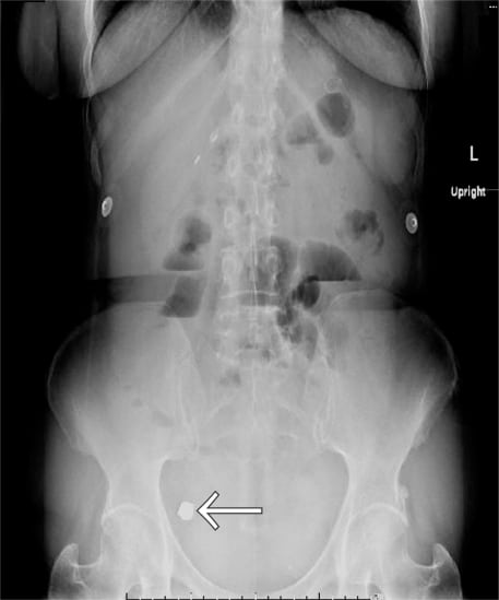
The complete blood count included a white count of 10,300 cells/mcL and hemoglobin of 13.7 g/dL. Electrolytes, hepatic function panel, lipase, renal function, and urinalysis were normal. An acute abdominal series showed multiple air fluid levels, distended small bowel, and a retained capsule endoscope in the terminal ileum consistent with a small bowel obstruction (Figure 1).
A nasogastric tube was placed and the patient was admitted. The patient was treated with IV steroids and antibiotics. The primary team performed a diatrizoate (Gastrografin) upper GI series with small bowel follow-through 12 hours after admission. Delayed films three hours later showed that the capsule had passed into the colon. Review of the endoscopic video images revealed erythema, granularity of the mucosa, friability, and edema of the small bowel, consistent with the diagnosis of Crohn’s disease.
DISCUSSION
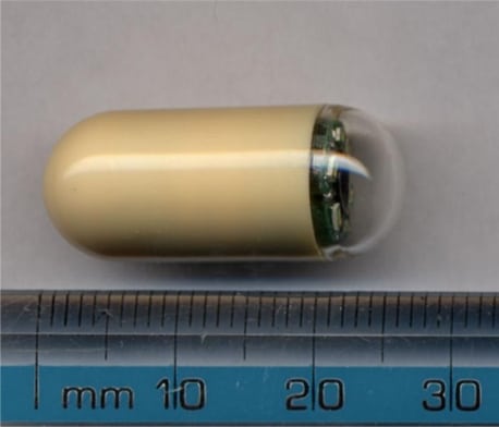
After its approval by the Food and Drug Administration in 2001, capsule endoscopy has gained widespread acceptance as an important imaging modality for small bowel pathology. Included in the 1 cm x 2.5 cm ingestible endoscope are a wireless video camera, illumination system, batteries, and an image transmitter (Figure 2). Images are recorded by a sensing system worn on the patient’s trunk, which is removed for image download approximately eight hours after capsule ingestion. Seventy-five percent of capsules enter the colon during the eight-hour acquisition time.1Subsequently, the disposable plastic-covered capsule is excreted in the stool 10–48 hours later.2,3
In preparation for capsule endoscopy, a patient typically fasts or maintains a clear liquid diet for 10–24 hours prior to the procedure. Several bowel preparation regimens (polyethylene glycol, simethicone, and/or sodium phosphate) have been studied, but there is no current consensus for whether bowel preparation is necessary or beneficial. Some clinicians administer promotility agents, including erythromycin and metoclopramide, during capsule passage. Evidence is controversial to support their use.4
Indications for capsule endoscopy include obscure GI bleeding, suspected Crohn’s disease, malabsorptive syndrome, or small intestinal tumors, and surveillance for patients with polyposis syndromes. The list of relative contraindications includes patients with known or suspected GI obstruction, stricture, fistula, or motility disorders, patients with cardiac pacemakers or other implanted electro-medical devices, dysphagia, and pregnancy.2,5
One of the common risks of the procedure is capsule retention. Capsule retention is defined as a capsule remaining in the gastrointestinal system for longer than two weeks, requiring directed medical, endoscopic, or surgical intervention to retrieve or pass the capsule.1 The most common causes of retention are Crohn’s disease, strictures caused by chronic non-steroidal anti-inflammatory drug (NSAID) use, small-bowel tumors, radiation enteritis, and surgical anastomotic strictures. The incidence of capsule retention ranges from 0% to 13%, with the most common site of detainment in the ileum, similar to our patient.1,6 Capsule retention occurs frequently in patients with known Crohn’s disease, 13% in one large study.7 Capsule retention rarely causes symptoms but can potentially cause small bowel obstruction or perforation.8–12
Asymptomatic capsule retention may be treated with medical, endoscopic, or surgical intervention.1,6 Capsule retrieval is not emergent in asymptomatic retainers. In one large study, early elective laparotomy or double-balloon endoscopy was performed to retrieve the capsule and treat the underlying cause for the stricture (small-bowel tumor, Crohn’s disease, ischemic bowel) in 34% of patients with asymptomatic capsule retention. The remainder (66%) underwent medical therapy for their underlying disease. Spontaneous capsule passage occurred in 52% of the medical therapy group. The rest underwent laparotomy for emergent obstructive symptoms (24%) and failure of medical treatment (24%).6 Medical therapies include steroids, infliximab, prokinetics, or cathartics. Their efficacy is controversial. The longest reported case of capsule retention is 2.5 years.1
Small bowel obstruction and perforation are rare but serious results of capsule retention. They may occur as early as several hours after capsule ingestion or after a prolonged period of capsule retention.8,9 Patients with abdominal pain, distension, and nausea after capsule endoscopy should be evaluated for impaction and small bowel obstruction or perforation. Diagnosis may be aided by interpretation of capsule images. There may be evidence of an obstructing lesion, repetitive images of the same area of mucosa, or inability to visualize the colon. Abdominal radiography or CT will typically demonstrate a radioopaque density in the small bowel.1
Cases of capsule retention with signs and symptoms of small bowel obstruction or perforation are managed operatively or non-operatively, depending on the clinical scenario. Non-surgical interventions include enemas and enteroscopy with snare retrieval.8,9 Surgical interventions may be open or laparoscopic with capsule recovery via enterectomy or intraoperative manual bowel manipulation and digital rectal retrieval.8,11,12 Surgical intervention can remove both the obstructing lesion and the retained capsule. This case report describes successful disimpaction with IV steroids and diatrizoate upper GI series with small bowel follow-through.
Of special note, patients with suspected capsule retention should not undergo magnetic resonance imaging.2 Risk of capsule non-passage should be assessed in any patient who has undergone capsule endoscopy in the previous three years.
Footnotes
Supervising Section Editor: Eric R. Snoey, MD
Submission history: Submitted July 24, 2009; Revision Received September 6, 2009; Accepted October 25, 2009
Full text available through open access at http://escholarship.org/uc/uciem_westjem
Address for Correspondence: Megan Boysen, MD, Department of Emergency Medicine, University of California, Irvine Medical Center, 101 The City Drive, Rte. 128-01, Orange, CA 92868
Email: mboysen@uci.edu
Conflicts of Interest: By the WestJEM article submission agreement, all authors are required to disclose all affiliations, funding sources, and financial or management relationships that could be perceived as potential sources of bias. The authors disclosed none.
REFERENCES
1. Cave D, Legnani P, de Ranchis R, et al. ICCE consensus for capsule retention. Endoscopy.2005;37:1065–7. [PubMed]
2. Mishkin DS, Chuttani R, Croffie J, et al. ASGE Technology Status Evaluation Report: wireless capsule endoscopy. Gastrointest Endosc. 2006;63(4):539–45. [PubMed]
3. Ali A, Santisi JM, Vargo J. Video capsule endoscopy: a voyage beyond the end of the scope. Cleve Clin J Med. 2004;71(5):415–25. [PubMed]
4. Mergener K, Ponchon T, Gralnek I, et al. Literature review and recommendations for clinical application of small-bowel capsule endoscopy, based on a panel discussion by international experts. Consensus statements for small-bowel capsule endoscopy, 2006/2007. Endoscopy. 2007;39:895–909. [PubMed]
5. Saurin JC. Capsule endoscopy. Endoscopy. 2007;39:986–91. [PubMed]
6. Cheon JH, Kim YS, Lee IS, et al. Can we predict spontaneous capsule passage after retention? A nationwide study to evaluate the incidence and clinical outcomes of capsule retention. Endoscopy.2007;39:1046–52. [PubMed]
7. Cheifetz AS, Korbluth AA, Legani P, et al. The risk of retention of the capsule endoscope in patients with known or suspected crohn’s disease. Am J Gastroenterol. 2006;101:2218–22. [PubMed]
8. Lin OS, Brandabur JJ, Schembre DB, et al. Acute symptomatic small bowel obstruction due to capsule impaction. Gastrointest Endosc. 2007;65(4):725–8. [PubMed]
9. Levsky JM, Milikow DL, Rozenblit AM, et al. Small bowel obstruction due to an impacted endoscopy capsule. Abdom Imaging. 2008;33(5):579–81. [PubMed]
10. Yu WK, Yang RD. M2A video capsule lodged in the Meckel’s divertiulum. Gastrointest Endosc.2006;63:1071–2. [PubMed]
11. Dominguez EP, Choi Y, Raijman IL, et al. Laparoscopic approach for the retreival of retained video capsule endoscopy. JLSL. 2006;10:496–8.
12. Ikorucu O, Tascilar O, Emre AU, et al. Small-bowel obstruction secondary to a wireless capsule enteroscopy: extraction of the capsule without enterotomy. Endoscopy. 2007;39:e286–87.[PubMed]