
| Author | Affiliation |
|---|---|
| Thomas Catron, MD | Alameda County Medical Center, Highland General Hospital, Department of Emergency Medicine |
| H. Gene Hern, MD, MS | Alameda County Medical Center, Highland General Hospital, Department of Emergency Medicine |
INTRODUCTION
Herpes zoster is a common diagnosis in the emergency department (ED). Caused by the reactivation of the varicellazoster virus (VZV), zoster usually presents as a painful dermatomal rash. In addition to skin or mucosal involvement, VZV reactivation commonly affects the ophthalmic division of the trigeminal nerve and subsequently the eye. This manifestation is termed herpes zoster ophthalmicus (HZO). HZO is considered an ophthalmologic emergency, as sequelae often include severe chronic pain and vision loss. In order to ensure proper follow up and to minimize morbidity, the accurate and timely diagnosis of HZO in the ED is extremely important. While typically an easy diagnosis to make based on history and skin findings, occasionally HZO presents as an isolated ophthalmologic process that is difficult to distinguish from other more benign causes of a red eye.
CASE REPORT
A 64-year-old Chinese man presented to the ED with five days of increasingly blurry vision in his right eye. The blurring was associated with mild photophobia and a right-sided headache which he described as a burning pain over his forehead. The patient did not wear corrective lenses and denied trauma, chemical exposure, discharge, foreign body sensation, sick contacts, or history of similar problems. Review of systems was positive for approximately one week of malaise, myalgia, and subjective fevers. The patient denied sore throat, rhinorrhea, hearing changes, rashes, or any other concerns. The patient did not know if he had ever had chicken pox or cold sores, and he denied any past medical problems, surgeries, allergies, or medications.
Physical exam revealed a well appearing male in no distress with vitals as follows: heart rate at 72 beats/minute, blood pressure at 145/84 mm Hg, respirations at 14 breaths/minute, oral temperature at 36.8°C and room air oxygen saturation at 97%. HENT exam showed no obvious rashes or lesions; however, the patient had mild hyperesthesia over his right forehead. He had normal ear canals, tympanic membranes, nares, and oropharynx. The patient had no cervical or auricular lymphadenopathy.
Ophthalmologic exam was significant for slight right-sided conjunctival irritation with no exudates or obvious corneal scarring. The patient had normal lids and consensual pupillary reflexes with mild discomfort on ipsilateral and contralateral pupillary testing. Extra-ocular motions were intact with no diplopia. Visual acuity was 20/20 in the left eye, 20/80 in the right, and 20/20 in both. Visual fields were normal and symmetric. Fundoscopic exam revealed normal appearing eye grounds with no evidence of hemorrhage, vascular occlusion, papilledema, or retinal detachment. Intraocular pressures were normal bilaterally. Anterior chamber slit lamp exam showed no corneal ulceration or cell and flare. Fluorescein staining yielded a 7mm branched dendritic corneal lesion (Figure 1). The remainder of his exam was unremarkable.
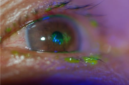
Dendritic fluorescein uptake from Herpes zoster opthalmicus.
The patient was started on oral acyclovir and a topical cycloplegic. Ophthalmology was consulted, and the patient was discharged to ophthalmology clinic, where the ED findings were confirmed. In clinic the patient was started on topical steroids in addition to antivirals and cycloplegics, and was discharged home with early follow up by ophthalmology.
DISCUSSION
Varicella-zoster virus manifests itself as two distinct syndromes in humans. During primary infection, the virus causes chicken pox. After the initial infection, VZV remains latent in the dorsal root ganglia of sensory neurons, possibly reappearing as herpes zoster later in the patient’s life.
In the United States, VZV primary infection rate reaches nearly 100% by 60 years of age; however, this rate will likely decrease with the widespread usage of the VZV vaccine. The lifetime risk of varicella zoster is between 10–20% in patients who have had chicken pox. Risks for reactivation include any decline in the T-cell mediated immune response including that caused by normal aging, HIV/AIDS, and immunosuppressive medications.
Herpes zoster ophthalmicus is a relatively common presentation of zoster. By definition HZO is reactivation of VZV in the ophthalmic division of the trigeminal nerve (V1), and accounts for 10–25% of all herpes zoster cases.1 While HZO does not necessarily affect the structures of the eye, many of the acute and long-term complications associated with the disease are the result of direct viral toxicity to the eye or the ensuing inflammatory response within the eye. It is thought that approximately 50% of those diagnosed with HZO will develop complications. Many of these poor outcomes can be prevented or ameliorated with early recognition, treatment, and referral.
Classically, HZO begins with flu-like symptoms including fever, myalgia, and malaise for approximately one week. Typically, patients then develop a painful unilateral dermatomal rash in the distribution of one or more branches of V1: supraorbital, lacrimal, and nasocilliary. The skin manifestations usually begin as an erythematous macular rash, progressing over several days into papules, vesicles, and then pustules. These eventually rupture and scab, and in immunocompetent individuals will resolve over the course of two to three weeks. In about 60% of cases patients will complain of a painful dermatomal prodrome prior to the development of any rash. Ocular involvement is not invariable in HZO; however, in patients with nasocilliary nerve involvement (Hutchinson’s sign) some case series indicate 100% go on to develop eye pathology.2Approximately one third of those without nasocilliary involvement will eventually develop eye manifestations.3 Conversely, in a small subset of patients (such as our patient), ocular symptoms will predominate.4
Physical exam should include a thorough ophthalmologic exam including external inspection, visual acuity, visual fields, extra ocular movements, pupillary response, funduscopy, intraocular pressure, anterior chamber slit lamp exam, and corneal exam with and without staining. As in our patient, the differential diagnosis includes a broad range of pathology: herpes simplex keratitis, other viral or bacterial conjunctivitis, uveitis, glaucoma, trauma, chemical exposure, vascular occlusion, migraine, cluster headache, trigeminal neuritis, optic neuritis, vasculitis, and others. If the classic rash is present, a brief exam will limit this differential; however, as in our patient, this would not be adequate to diagnose a patient presenting with purely ocular involvement. Classic ocular involvement is typified by dendritic or punctate keratitis (Figure 1). This pattern of infection occurs in approximately 65% of patients with HZO;4 however, other eye findings are more frequent and range from simple conjunctivitis to retinal necrosis and detachment. Any structure in the eye may be involved.4
Diagnostic testing is rarely indicated, as diagnosis can almost always be made by a combination of history and physical. It is possible to use a Tzanck smear or Wright stain to determine whether lesions contain herpes-type virus (though these will not differentiate between VZV and other herpes viruses). Viral culture, direct immunoflourescence assay, or PCR may also be used to confirm the diagnosis.
In the ED treatment consists of local wound care, pain control, initiation of antiviral medication, and antibiotics if needed. Acyclovir and other similar antivirals have been shown to significantly decrease adverse outcomes related to HZO if started within 72 hours of initial symptoms.5,6,7,8 Studies report reduced pain during the outbreak, reduced likelihood of postherpetic neuralgia, increased rate of skin healing, decreased duration of viral shedding, and decreased incidence of corneal involvement. It is not proven that the patient accrues these benefits if the medications are started after the 72-hour window, but given the extremely low side-effect profile of the drugs and the serious sequelae of complications, most physicians recommend administering antivirals during the first seven to 10 days of symptoms.
Steroids (topical and systemic) may also play a role in the treatment of HZO. In some studies systemic steroids have been shown to speed skin healing and to decrease initial pain; however, there have been no definitive studies showing reduced long-term incidence of post-herpetic neuralgia or ocular complications.9,10 Likewise, topical steroids may be helpful in the initial management of pain due to uveitis or scleritis; however, they have a number of serious side effects and potential complications and should never be used without concomitant anti-viral treatment.4 Always obtain ophthalmologic consultation prior to starting steroid treatment.
Oral opiate and nonsteroidal anti-inflammatory medications are frequently indicated for pain and may be augmented by the use of cycloplegics in patients who display features of iritis.
In otherwise healthy individuals with minimal eye involvement, most sources suggest outpatient treatment with seven to 10 days of oral acyclovir at a dose of 800mg five times and famciclovir) offer equivalent benefits and have reduced frequency of dosing, which may improve patient compliance. In high risk cases, admission for IV acyclovir is indicated. Admission is recommended for those with known immunodeficiency, patients on immunosuppressive medications, involvement of multiple dermatomes (which may indicate immunosuppression), retinal involvement, corneal ulceration, or serious bacterial superinfection. All patients with the possible diagnosis of HZO require ophthalmologic consultation prior to discharge from the ED in order to ensure full evaluation for more serious complications. For patients eventually discharged home, early ophthalmologic follow up is mandatory.
CONCLUSION
HZO is a potentially serious reactivation of VZV in the distribution of the ophthalmic division of the trigeminal nerve. Usually this entity presents with classic findings that make diagnosis simple; however, as was the case with our patient, HZO may easily be confused with more common and benign eye pathology. Full ophthalmologic exam is warranted in the patient with decreased vision or a red eye. Once the diagnosis of HZO is entertained, appropriate antiviral and adjunctive therapy should be initiated and ophthalmology consultation should be requested for evaluation and treatment of common complications.
Footnotes
Supervising Section Editor: Eric R. Snoey, MD
Submission history: Submitted November 15, 2007; Revision Received None; Accepted February 22, 2008.
Full text available through open access at http://escholarship.org/uc/uciem_westjem
Address for Correspondence: H. Gene Hern, Jr. MD, MS, Department of Emergency Medicine, Alameda County Medical Center, Highland General Hospital, 1411 E 31st St, Oakland, CA 94602-1018
Email: gene_hern@yahoo.com
Conflicts of Interest: By the WestJEM article submission agreement, all authors are required to disclose all affiliations, funding sources, and financial or management relationships that could be perceived as potential sources of bias. The authors disclosed none.
REFERENCES
1. Ragozzino MW, Melton LJ, 3d, Kurland LT, Chu CP, Perry HO. Population-based study of herpes zoster and its sequelae. Medicine. 1982;61:310–316. [PubMed]
2. Zaal MJ, Völker-Dieben HJ, D’Amaro J. Prognostic value of Hutchinson’s sign in acute herpes zoster ophthalmicus. Graefes Arch Clin Exp Ophthalmol. 2003;241:187–191.[PubMed]
3. Harding SP, Lipton JR, Wells JC. Natural history of herpes zoster ophthalmicus: predictors of postherpetic neuralgia and ocular involvement. Br J Ophthalmol.1987;71:353–358. [PMC free article] [PubMed]
4. Shaikh S, Ta CN. Evaluation and management of herpes zoster ophthalmicus. Am Fam Physician. 2002;66:1723–1730. [PubMed]
5. Severson EA, Baratz KH, Hodge DO, Burke JP. Herpes zoster ophthalmicus in olmsted county, Minnesota: have systemic antivirals made a difference? Arch Ophthalmol.2003;121:386–390. [PubMed]
6. Morton P, Thomson AN. Oral acyclovir in the treatment of herpes zoster in general practice. N Z Med J. 1989;102:93–95. [PubMed]
7. Huff JC, Bean B, Balfour HH, Jr, Laskin OL, Connor JD, Corey L, et al. Therapy of herpes zoster with oral acyclovir. Am J Med. 1988;85:84–89. [PubMed]
8. McGill J, Chapman C, Mahakasingam M. Acyclovir therapy in herpes zoster infection. A practical guide. Trans Ophthalmol Soc U K. 1983;103:111–114. [PubMed]
9. Wood MJ, Johnson RW, McKendrick MW, et al. A randomized trial of acyclovir for 7 days or 21 days with and without prednisolone for treatment of acute herpes zoster. N Engl J Med. 1994;330:896–900. [PubMed]
10. Whitley RJ, Weiss H, Gnann JW, et al. Acyclovir with and without prednisone for the treatment of herpes zoster. A randomized, placebo-controlled trial. The National Institute of Allergy and Infectious Diseases Collaborative Antiviral Study Group. Ann Intern Med.1996;125:376–383. [PubMed]