| Author | Affiliation |
|---|---|
| Sahin Bozok, MD | Rize University Faculty of Medicine, Rize Research and Training Hospital, Rize, Turkey |
| Özcan Yavaşi, MD | Rize State Hospital, Rize, Turkey |
| Gokhan Ilhan, MD | Rize University Faculty of Medicine, Rize Research and Training Hospital, Rize, Turkey |
| Ali Gurbuz, MD | Katip Celebi University Faculty of Medicine, Izmir Ataturk Research and Training Hospital, Izmir, Turkey |
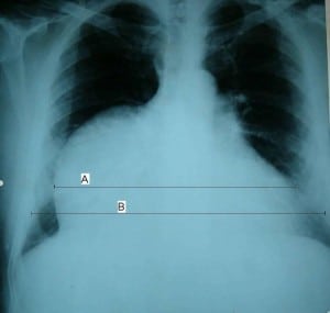
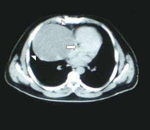
A 29-year-old man with sudden onset of dyspnea and chest pain with impairment of the general status after falling down from five meters was transferred to our emergency department. He was completely asymptomatic before the injury, but hypotensive (80/50 mmHg) and tachycardic (112 beats/minute) after the injury. Chest radiograph revealed a bulging cardiac silhouette on the right paracardiac region with an increased cardiothoracic ratio of 70% (Figure 1a). Computed tomography revealed a giant cystic mass with a diameter of 10 cm making cardiac compression (Figure 1b). On transthoracic echocardiography, a huge mass neighboring the right atrium with 15×10 cm dimensions was seen. The mass was outside of the pericardial cavity. The left ventricular ejection fraction (LVEF) was 40%. The patient was taken to urgent operation. A huge unruptured cystic mass, which had no connection with the pericardium, was easily resected. Histopathologically, it was reported as a cystic thymoma. On the third postoperative day, echocardiography showed normal cardiac functions with a LVEF of 60%. The patient was discharged without any complications and asymptomatic after three months following surgery.

Posteroanterior chest radiogram showing increased cardiothoracic ratio (brackets). The cardiac diameter (a) is 70% of the transthoracic diameter (b).

Contrast-enhanced computed tomography showing a large cystic mass (arrowheads)causing cardiac compression (arrows).
Thymomas are the most common anterior mediastinal masses in adults, accounting for 15% all mediastinal tumors and 30 to 50% of them are asymptomatic.1,2 It is reported that 40% of thymomas are characterized by cystic degeneration.3
In the present case, the huge cystic thymoma was present in the anterior mediastinum for many years without any symptoms. It was firstly misdiagnosed as pericardial tamponade because of a history of injury. A giant cyst formation compressing the heart became obvious after imaging. Although a definitive diagnosis can be obtained by the histopathological examination of the operative material, cystic thymoma should be considered in patients with anterior mediastinal cystic masses.
Footnotes
Supervising Section Editor: Sean O. Henderson, MD
Submission history: Submitted December 10, 2011; Accepted March 5, 2012
Full text available through open access at http://escholarship.org/uc/uciem_westjem
DOI: 10.5811/westjem.2012.3.11562
Address for Correspondence: Özcan YAVAŞi, MD, Rize State Hospital, Department of Emergency Medicine, Eminettin Mahallesi, Ataturk Caddesi, 53100 Rize, Turkey
Email: ozcanyavasi@yahoo.com.tr
Conflicts of Interest: By the WestJEM article submission agreement, all authors are required to disclose all affiliations, funding sources, and financial or management relationships that could be perceived as potential sources of bias. The authors disclosed none.
REFERENCES
1. Strollo DC, De Christenson MLR, Jett JR. Primary mediastinal tumors. Part 1. Tumors of the anterior mediastinum. Chest. 1997;112:511–22. [PubMed]
2. Morgenthaler TI, Brown LR, Colby TV, et al. Thymoma. Mayo Clin Proc.1993;68:1110–23. [PubMed]
3. Suster S, Rosai J. Cystic thymomas: a clinicopathologic study of ten cases. Cancer.1992;69:92–7. [PubMed]


