
| Author | Affiliation |
|---|---|
| Stacey L. Poznanski, DO | University of Wisconsin School of Medicine and Public Health, Division of Emergency Medicine, Madison, WI |
| Gerard S Doyle, MD, MPH | University of Wisconsin School of Medicine and Public Health, Division of Emergency Medicine, Madison, WI |
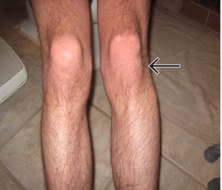
A 17-year-old man presented with acute left lateral knee pain after “twisting” his knee during a soccer scrimmage. He denied trauma and prior injury to that knee. He was unable to bear weight but denied neurovascular symptoms. Examination revealed a prominence deformity and significant tenderness over the lateral knee distal to the joint line, in the region of the fibular head. During his emergency department (ED) evaluation, he had intact peroneal nerve function. A picture of his legs is shown in Figure 1.
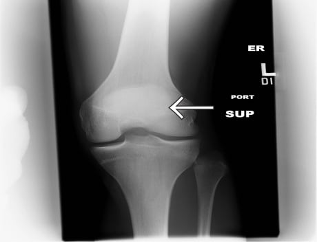
Proximal tibiofibular joint (PTFJ) dislocation is an unusual injury, occurring in less than 1% of knee injuries. It has been reported in soccer, rugby, and football players, ballet dancers, parachutists and snowboarders.1,2 It typically occurs when the knee is slightly flexed and the foot is rotated and plantar flexed.2 Radiographic findings can be subtle, and diagnosis is fostered when comparison films are obtained. When in doubt, computed tomography should be obtained.3 Our radiographs demonstrated widening of the PTFJ of the left knee; comparison to the right (uninjured) knee confirms the diagnosis (Figure 2).
We were unable to reduce the dislocation despite moderate sedation with etomidate. The patient was also seen by orthopedic surgery in the ED. At follow-up two days later, he was noted to have mildly decreased sensation in the peroneal nerve distribution. He was scheduled for surgery, but prior to repair he slipped and felt a “pop.” Pre-operative radiographs revealed that the dislocation had spontaneously reduced.
Footnotes
Supervising Section Editor: Sean Henderson, MD
Submission history: Submitted January 8, 2010; Revision Received January 10, 2010; Accepted January 11, 2010
Full text available through open access at http://escholarship.org/uc/uciem_westjem
Address for Correspondence: Gerard S Doyle, MD MPH, Division of Emergency Medicine, University of Wisconsin School of Medicine and Public Health, 600 Highland Drive, Madison, WI 53792
Email: gsdoyle@medicine.wisc.edu
Conflicts of Interest: By the WestJEM article submission agreement, all authors are required to disclose all affiliations, funding sources, and financial or management relationships that could be perceived as potential sources of bias. The authors disclosed none.
REFERENCES
1. Ahmad R, Case R. Dislocation of the fibular head in an unusual sports injury: a case report. J Med Case Reports. 2008;(2):158–60. [PMC free article] [PubMed]
2. Horan J, Quin G. Proximal tibiofibular dislocation. Emerg Med J. 2006;23(5):e33–4.[PMC free article] [PubMed]
3. Keogh P, Masterson E, Murphy B, et al. The role of radiography and computed tomography in the diagnosis of acute dislocation of the proximal tibiofibular joint. Br J Radiol. 1993;66(782):108–11.[PubMed]