| Author | Affiliation |
|---|---|
| Philippe Hantson, MD, PhD | Université Catholique de Louvain, Brussels, Belgium |
| Thierry Duprez, MD | Université Catholique de Louvain, Brussels, Belgium |
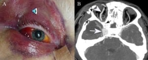
A 54-year-old man presented to the emergency department with a Glasgow Coma Score of 8 after throwing himself out of a window. Right periorbital and conjunctival blood infiltration were noted, with complete ophthalmoplegia and loss of the pupillary light reflex. On the third day, a systolic bruit became audible over the right eyeball [(Figure 1A) Click on image for audio]. The CT with contrast (Figure 1B) demonstrated a wide unilateral enlargement of the right cavernous sinus (white arrow) and of the superior ophthalmic vein (arrowhead), together with multiple bone disruptions, including the sphenoid bone (black arrows). This was consistent with the diagnosis of traumatic carotid cavernous fistula (TCCF). A TCCF arises from direct traumatization of the internal carotid artery within its cavernous course. It is a relatively rare complication of head trauma, with a global occurrence rate ranging from 0.17 to 1.01%.1Usually TCCF are found after fractures of the middle third portion of the facial bone. When they are of high-flow type, they develop within a short period after injury.

The diagnosis of TCCF is difficult in comatose patients. Complete ophthalmoplegia, with an ocular bruit, should prompt careful analysis of the head CT scan. High-flow TCCFs should be treated as a relative emergency. They may cause secondary neurological deficits due to flow steal from the intracranial carotid artery or ophthalmologic complications secondary to venous congestion. Endoarterial detachable balloon embolization is the treatment of choice.2
Footnotes
Supervising Section Editor: Kurt R. Denninghoff, MD
Submission history: Submitted February 27, 2008; Revision Received June 30, 2008; Accepted September 12, 2008.
Full text available through open access at http://escholarship.org/uc/uciem_westjem
Address for Correspondence: Philippe Hantson, MD, PhD. Cliniques St. Luc, Avenue Hippocrate 10, Brussels 1200, Belgium
Email: philippe.hantson@uclouvain.be
Conflicts of Interest: By the WestJEM article submission agreement, all authors are required to disclose all affiliations, funding sources, and financial or management relationships that could be perceived as potential sources of bias. The authors disclosed none.
REFERENCES
1. Takenoshita Y, Hasuo K, Matsushima T, Oka M. Carotid-cavernous sinus fistula accompanying facial trauma. Report of a case with a review of the literature. J Craniomaxillofac Surg. 1990;18:41–45. [PubMed]
2. Higashida RT, Halbach VV, Tsai FY, Norman D, Pribran HF, Mehringer CM. Interventional neurovascular treatment of traumatic carotid and vertebral lesions: results in 234 cases. Am J Roentgenol. 1989;153:577–582. [PubMed]


