
| Author | Affiliation |
|---|---|
| Tommaso Bartalena | Poliambulatorio Privato Zappi Bartalena Centro Medico Diagnostico, Imola (BO), Italy |
| Maria Francesca Rinaldi | Radiologia III – Pol.S.Orsola-Malpighi, Bologna, Italy |
| Carlo De Luca | Radiologia d’Urgenza – Pol.S.Orsola-Malpighi, Bologna, Italy |
| Eugenio Rimondi | Radiologia – Istituto Ortopedico Rizzoli, Bologna, Italy |
A 68-year-old male presented to the emergency department with anterior right knee pain after falling at home and directly hitting his patella on the stepladder edge while he was going upstairs. Physical examination revealed infrapatellar region painful swelling and impaired knee extension. Knee radiographs showed no fracture; however, the lateral view revealed a high riding patella, concerning for a patellar tendon lesion (Figure 1). Ultrasounds confirmed radiographic suspect showing complete patellar tendon rupture (Figure 2) with proximal retraction of the patella.
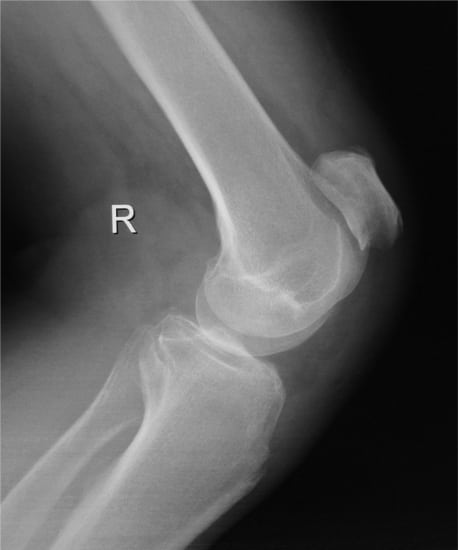
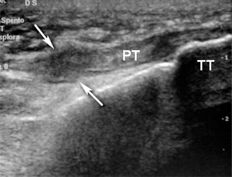
In a normal knee the ratio between the patellar tendon length and the diagonal length of the patella (Insall-Salvati ratio) should be approximatiely one, while this patient had a ratio of 2.2.1 Differential diagnosis of patella alta includes patellar tendon rupture, subluxation of the patella, Sinding-Larsen-Johansson disease and joint effusion.2 The patellar tendon composes the knee extensor mechanism together with the quadriceps tendon. Patellar tendon ruptures are less common than quadriceps tendon ruptures and are typically seen in those under 40 years of age. The injury may be either direct or indirect. The more common indirect mechanism results from forced flexion when the quadriceps is contracted. The direct mechanism, which is less common, is the result of a violent impact against a taut quadriceps tendon. Most ruptured patellar tendons occur over chronically degenerated tendons. Steroid injections are thought to predispose to rupture. Other predisposing factors include tendon calcifications, arthritis, collagen disorders, fatty tendon degeneration, and metabolic disorders.3 Patella alta on knee radiographs in a trauma setting should prompt further imaging to rule out patellar tendon rupture. Ultrasound is a very cost-effective technique to evaluate knee tendons and ligaments; it is a rapidly available imaging modality and represents the easiest and quickest method to diagnose a tear and determine its site and extent.4 The patellar tendon is imaged with longitudinal scans using high-frequency linear transducers; it normally appears as a continuous well-defined hyperechoic fibrillar structure bridging the patella and the tibial tuberosity. while tears appear as hypoechoic areas of interruption of the fibrillar pattern.
Footnotes
Supervising Section Editor: Sean Henderson, MD
Submission history: Submitted June 12, 2009; Revision Received June 17, 2009; Accepted June 17, 2009
Full text available through open access at http://escholarship.org/uc/uciem_westjem
Address for Correspondence: Dr. Tommaso Bartalena, Poliambulatorio Privato Zappi Bartalena Centro Medico Diagnostico, Via Cogne 4 – 40026 – Imola (BO), Italy
E-mail: t.bartalena@email.it
Conflicts of Interest: By the WestJEM article submission agreement, all authors are required to disclose all affiliations, funding sources, and financial or management relationships that could be perceived as potential sources of bias. The authors disclosed none.
REFERENCES
1. Insall J, Salvati E. Patella position in the normal knee joint. Radiology. 1971;101:101–4. [PubMed]
2. Taylor JAM, Resnick D. Skeletal Imaging – Atlas of the Spine and Extremities. 1st ed. Philadelphia, PA: Saunders, WB; 2000.
3. Simon RR, Sherman SC, Keonigknecth SJ. Emergency Orthopedics: The Extremities. 5th ed. New York, NY: McGraw-Hill Medical; 2007.
4. Matava MJ. Patellar tendon ruptures. J Am Acad Orthop Surg. 1996;4:287–96. [PubMed]