
| Author | Affiliation |
|---|---|
| Prerna Gupta, MD, DNB | Department of Obstetrics and Gynecology, India Institute of Medical Sciences, New Delhi, India |
| Ankur Gadodia, MD, DNB | Department of Radio-diagnosis, India Institute of Medical Sciences, New Delhi, India |
| Ashu Seith, MD | Department of Radio-diagnosis, India Institute of Medical Sciences, New Delhi, India |
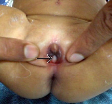
An 8-day-old female presented with abdominal distension, anuria and a translucent membrane protruding from the vagina. Clinical examination revealed a palpable abdomino-pelvic fluctuant mass lesion. On perineal examination, a translucent bulge was seen and increased in size when the neonate cried or when pressure was exerted on the abdominal mass (Figure 1). Differential diagnoses considered were imperforate hymen, urethral prolapse and prolapsed ureterocoele. Ultrasound of the abdomen revealed a large abdomino-pelvic cystic mass lesion containing mobile internal debris (Figure 2). Bilateral moderate hydronephrosis was also seen. Diagnosis of imperforate hymen causing hydrometrocolpos, anuria and bilateral hydronephrosis was made.

The bulging membrane was incised and drained 300 mL of fluid. The child improved symptomatically. Hydronephrosis resolved on follow-up ultrasonography. An imperforate hymen causing hydrometrocolpos, bilateral hydronephrosis and associated anuria/urinary retention is extremely rare in the neonatal period.1 Neonatal Hydrometrocolpos secondary to imperforate hymen usually presents soon after birth with a lower abdominal mass and a bulging introitus. Other reported presentations include acute renal failure, respiratory distress, sepsis, and intestinal obstruction.2 Sonographic findings include cystic abdomino-pelvic mass containing debris with posterior acoustic enhancement and hydrosalpinx.3 Hymenectomy through perineal approach is the mainstay of management.1
Footnotes
Supervising Section Editor: Rick McPheeters, MD
Submission history: Submitted February 6, 2010; Revision Received March 12, 2010; Accepted March 29, 2010
Full text available through open access at http://escholarship.org/uc/uciem_westjem
Address for Correspondence: Ashu Seith Bhalla, MD, Department of Radio-diagnosis, India Institute of Medical Sciences, New Delhi, India-110029
E-mail: ashubhalla1@yahoo.com
Conflicts of Interest: By the WestJEM article submission agreement, all authors are required to disclose all affiliations, funding sources, and financial or management relationships that could be perceived as potential sources of bias. The authors disclosed none.
REFERENCES
1. Nazir Z, Rizvi RM, Qureshi RN, et al. Congenital vaginal obstructions: varied presentation and outcome. Pediatr Surg Int. 2006;22:749–53. [PubMed]
2. El-Messidi A, Fleming NA. Congenital imperforate hymen and its life-threatening consequences in the neonatal period. J Pediatr Adolesc Gynecol. 2006;19:99–103. [PubMed]
3. Banerjee AK, Clarke O, MacDonald LM. Sonographic detection of neonatal hydrometrocolpos. Br J Radiol. 1992;65:268–71. [PubMed]