
| Author | Affiliation |
|---|---|
| Elizabeth S. Buyers, MD | Maine Medical Center, Department of Emergency Medicine, Portland, ME |
| Sara W. Nelson, MD | Maine Medical Center, Department of Emergency Medicine, Portland, ME |
| George L. Higgins, III, MD | Maine Medical Center, Department of Emergency Medicine, Portland, ME |
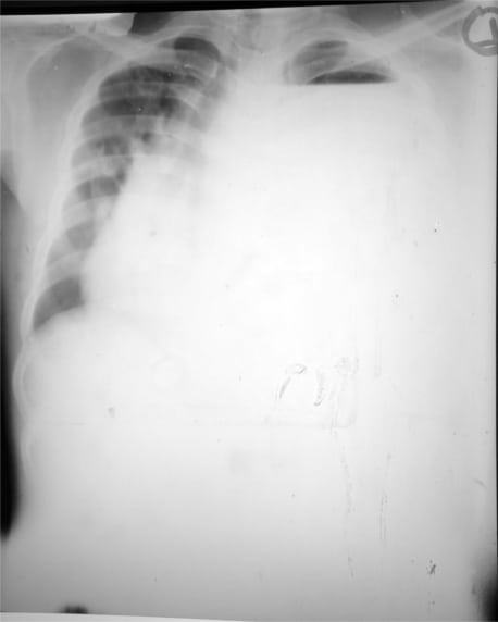
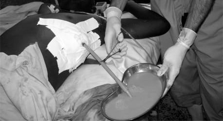
A 22-year-old man presented to a rural Ugandan clinic with three months of progressive dyspnea. He described a non-productive cough and subjective fevers and chills. He appears mildly dyspneic but is in no acute distress. His has a temperature of 37.7°C, pulse of 112 beats per minute, respiratory rate of 22 breaths per minute, blood pressure of 105/50 mmHg and an oxygen saturation of 93% on room air. Examination reveals absent breath sounds over the left chest. Chest radiograph demonstrates a massive fluid collection in his left hemi-thorax (Figure 1). Subsequent tube thoracostomy was productive of over three liters of purulent material (Figure 2). The patient tolerated the procedure without complication and was started on broad-spectrum antibiotics. AFB studies were eventually negative, but he was lost to follow-up.
Patients with massive empyema, although uncommon in this country, are likely to be more frequently encountered as international medicine experiences increase. The early goals of empyema therapy include evacuation of the purulent collection, sterilization of the pleural cavity, and lung re-expansion.1 Drainage requires aggressive management, such as large bore (at least 28 French) tube thoracostomy, with or without fibrinolytic therapy. The outer fibrin pleural peel of the empyema must be penetrated.
Complications from evacuating massive empyemas include re-expansion pulmonary edema (in adults from any cause and in children with malignant lymphoma), hemorrhage, secondary infection, pneumothorax and esophagopleural fistulas.2,3 To prevent these complications, sterile technique is required and image guidance by computed tomography and/or ultrasound may be useful. It is recommended that no more than 1500 mL of fluid be drained at one time or that the drainage be limited to no more than 500 mL/hour.4 Incomplete drainage may be attributed to pleural loculations or tube clogging, kinking or malposition.3 Debridement via video-assisted thoracoscopy or open thoracotomy may be required.5
Footnotes
Supervising Section Editor: Sean O. Henderson, MD
Submission history: Submitted March 1, 2010; Revision Received March 23, 2010; Accepted March 29, 2010
Full text available through open access at http://escholarship.org/uc/uciem_westjem
Address for Correspondence: George L. Higgins, III, MD, Director of Research, Department of Emergency Medicine, Maine Medical Center, 47 Bramhall Street, Portland, ME 04102
Email: higgig@mmc.org
Conflicts of Interest: By the WestJEM article submission agreement, all authors are required to disclose all affiliations, funding sources, and financial or management relationships that could be perceived as potential sources of bias. The authors disclosed none.
REFERENCES
1. Jaffe A, Balfour-Lynn IM. Management of empyema in children. Pediatric Pulmono. 2005;40:148–56.
2. Trapnell DH, Thurston JG. Unilateral pulmonary edema after pleural aspiration. Lancet.1970;1:1367–9. [PubMed]
3. Stark DD, Federle MP, Goodman PC. CT and radiographic assessment of tube thoracostomy. AJR.1983;141:253–8. [PubMed]
4. Balfour-Lynn IM, Abrahamson E, Cohen G, et al. BTS guidelines for the management of pleural infection in children. Thorax. 2005;60(Suppl 1):i1–i21. [PMC free article] [PubMed]
5. Avansino JR, Goldman B, Sawin RS, et al. Primary operative versus nonoperative therapy for pediatric empyema: a meta-analysis. Pediatrics. 2005;115:1652–9. [PubMed]