| Author | Affiliation |
|---|---|
| Paul Geukens, MD | Universite catholique de Louvain, Cliniques St Luc, Intensive Care, Brussels, Belgium |
| Thierry Duprez, MD | Universite catholique de Louvain, Cliniques St Luc, Neuroradiology, Brussels, Belgium |
| Philippe Hantson, MD, PhD | Universite catholique de Louvain, Cliniques St Luc, Intensive Care, Brussels, Belgium |
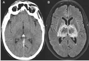
A 35-year-old woman, without previous medical history except oral contraception, presented with sudden onset of stupor and clonic perseveration in the upper limbs. She was aphasic, but communicated by vertical movements of the head. Unenhanced brain computed tomography (CT) demonstrated hyperintensity of the straight sinus and hypo-intense areas within both thalami (Figure 1A) leading to a diagnosis of cerebral venous thrombosis (CVT). Despite anticoagulation therapy, the patient deteriorated with a Glasgow Coma Score at 7. A brain magnetic resonance imaging (MRI) obtained two days later confirmed extensive acute venous thrombosis involving the straight sinus, the right tentorial sinus, the left proximal tentorial sinus and the vein of Galan. A bilateral thalamic venous infarction was confirmed (Figure 1B). Local thrombectomy followed by thrombolysis within the straight sinus was unsuccessful. A mild hemorrhagic transformation of the bithalamic infarction was observed on follow-up MRI. Neurological status slowly improved over time. At 3-month follow-up, she had no cognitive deficit and was able to walk spite of residual spasticity.

A. Unenhanced brain computed tomography. Strong hyperintensity within straight sinus due to the presence of an acute clot (arrow) together with subtle hypo-intensity within both thalamomesencephalic junctions. B. Magnetic resonance imaging with Fluid Attenuated Inversion Recovery (FLAIR) sequence. Hemorrhage within both thalami (central hyposignal intensity due to the presence of deoxyhemoglobin and marginal hyper signal intensity due to surrounding vasogenic edema). Thin residual clot within straight sinus (arrow) and moderate enlargement of lateral ventricles.
Bilateral infarctions of the thalami may be observed from either arterial or venous origin. They account for only 0.6% of all cerebral infarctions. Occlusion of the top of the basilar artery or of the so-called artery of Percheron – a developmental variant replacing the perforating medial thalamic arteries – is responsible for arterial ischemia. On venous side, the posterior group of thalamic veins drains into the straight sinus. A thrombosis of this vessel may also lead to a bilateral partial venous infarct due to the upstream overpressure.
The diagnosis of CVT is often challenging, as clinical symptoms are highly variable and unspecific.1 Good clinical recovery may be observed, even after severe and prolonged deficits at early acute stage of the disease course. Common risk factors for CVT, when identified, include pregnancy, early post-partum, oral contraception, hypercoagulability and infection.
Footnotes
Supervising Section Editor: Sean O. Henderson, MD
Submission history: Submitted March 30, 2012; Accepted April 09, 2012
Full text available through open access at http://escholarship.org/uc/uciem_westjem
DOI: 10.5811/westjem.2012.4.12250
Address for Correspondence: Philippe Hantson, MD, PhD. Universite catholique de Louvain, Cliniques St Luc, Intensive Care, Brussels, Belgium
Email: philippe.hantson@uclouvain.be
Conflicts of Interest: By the WestJEM article submission agreement, all authors are required to disclose all affiliations, funding sources, and financial or management relationships that could be perceived as potential sources of bias. The authors disclosed none.
REFERENCES
1. Gossner J, Larsen J, Knauth M. Bilateral thalamic infarction-a rare manifestation of dural sinus thrombosis. Clin Imaging. 2010;34:134–37. [PubMed]


