| Author | Affiliation |
| Scott Sullivan, MD | Madigan Army Medical Center, Department of Emergency Medicine, Tacoma, Washington |
| Kevin Maskell, MD | Madigan Army Medical Center, Department of Emergency Medicine, Tacoma, Washington |
| Tristan Knutson, MD | Madigan Army Medical Center, Department of Emergency Medicine, Tacoma, Washington |
Presentation
Diagnosis/Discussion
PRESENTATION
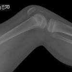
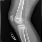
A 10 year-old male presented to the ED with knee pain after falling off his bicycle. He landed on his flexed knee with an audible “pop.” He could not extend his knee or walk. Physical examination revealed an effusion and high riding patella with a palpable inferior pole defect. He was neurovascularly intact, and the remaining examination of his lower extremity was unremarkable. His radiographs are shown in Figure 1.
DIAGNOSIS/DISCUSSION
Patellar Sleeve Fracture (PSF)
Patellar fracture and tendon rupture are uncommon occurrences in skeletally immature children. In this population, the most common form of this injury is the PSF, which should be considered in patients with an exam concerning for patellar tendon rupture.1 During secondary ossification, the patella is surrounded in a layer of protective cartilage. Forced loads on the contracted quadriceps with knee flexion results in an avulsion fracture. Since much of the fragment is unossified peripheral cartilage, these sleeve fractures are difficult to detect on radiographs. If there is clinical suspicion for PSF, magnetic resonance imaging can visualize this fragment.2 Identifying sleeve fractures is critical as prompt surgical correction is required for recovery of function.3 Delay may result in complications such as reduced knee flexion, ectopic bone formation, or avascular necrosis.3,4
Figure 1. This is the initial lateral view of the patient’s knee demonstrating patella alta and a 1.3cm ossific fragment representing an avulsion with partial rupture of the patellar sleeve.
This patient’s x-rays (Figure 1) reveal patella alta and a 1.3cm ossific fragment representing an avulsion with partial rupture of the patellar sleeve. He had an ORIF the next day. At 15 week follow up, his patellar tendon remained intact and he was progressing in physical therapy. His post-operative radiograph is displayed in Figure 2.
Figure 2. This is the post-surgical fixation lateral view of the patient’s knee demonstrating resolution of patella alta and fixation of the ossific fragment.
Footnotes
Supervising Section Editor: Sean O. Henderson, MD
Full text available through open access at http://escholarship.org/uc/uciem_westjem
Address for Correspondence: Scott Sullivan, MD, 3086 O’Brien St, DuPont, WA 98327. Email: scott.b.sullivan1@gmail.com.
Submission history: Submitted April 1, 2014; Revision received August 24, 2014; Accepted August 30, 2014
Conflicts of Interest: By the WestJEM article submission agreement, all authors are required to disclose all affiliations, funding sources and financial or management relationships that could be perceived as potential sources of bias. The authors disclosed none.
REFERENCES
- Dai LY, Zhang WM. Fractures of the patella in children. Knee Surg Sports Traumatol Arthrosc. 1999;7:243-245. [PubMed]
- Bates DG, Hresko MT, Jaramillo D. Patellar sleeve fracture: demonstration with MR imaging. Radiology. 1994;193(3):825-7. [PubMed]
- Guy SP, Marciniak JL, Tulwa N, et al. Bilateral sleeve fracture of the inferior poles of the patella in a healthy child: case report and review of the literature. Adv Orthop. 2011;2011:428614. [PubMed]
- Hunt DM, Somashekar N. A review of sleeve fractures of the patella in children. Knee. 2005;12(1):3-7. [PubMed]


