
| Author | Affiliation |
|---|---|
| Abdel-Rauf Zeina | Department of Radiology, Hillel Yaffe Medical Center, Hadera, Israel. Affiliated to Faculty of Medicine, Technion-Israel Institute of Technology, Haifa, Israel |
| Eiass Kassem | Pediatric Department, Hillel Yaffe Medical Center, Hadera, Israel. Affiliated to Faculty of Medicine, Technion-Israel Institute of Technology, Haifa, Israel |
| Adi Klein | Pediatric Department, Hillel Yaffe Medical Center, Hadera, Israel. Affiliated to Faculty of Medicine, Technion-Israel Institute of Technology, Haifa, Israel |
| Alicia Nachtigal | Department of Radiology, Hillel Yaffe Medical Center, Hadera, Israel. Affiliated to Faculty of Medicine, Technion-Israel Institute of Technology, Haifa, Israel |
A two-year-old girl presented with poor appetite, vomiting and decreased level of consciousness. Brain unenhanced computed tomography (CT) on admission demonstrated no hemorrhages but a hyperdense appearance of the left sigmoid sinus, left and right transverse sinus, and the superior sagittal sinus, consistent with the “cord sign” and representing acute thrombus (Figure 1a–c). Brain CT venography (obtained after administration of contrast agent) showed filling defects within the same cerebral veins corresponding to extensive sinus vein thrombosis (SVT) (Figure 1d–f).
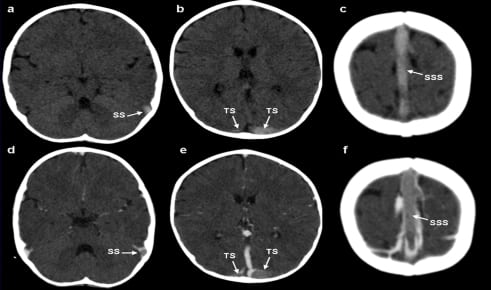
On unenhanced CT scan, thrombus appears hyperdense for the first 7–14 days.1 Its prevalence is variable and generally accepted to be an accurate sign when present.2 It is very important to diagnose this condition as early as possible so that specific treatment can be started. The cord sign (hyperdense thrombosed veins on unenhanced CT) and the empty-delta sign (a filling defect in the superior sagittal sinus on enhanced CT) are considered pathognomonic for SVT.
Footnotes
Supervising Section Editor: Sean Henderson, MD
Submission history: Submitted Janurary 9, 2010; Revision Janurary 11, 2010; Accepted January 19, 2010
Full text available through open access at http://escholarship.org/uc/uciem_westjem
Address for Correspondence: Abdel-Rauf Zeina, MD, Department of Radiology, Hillel Yaffe Medical Center, P.O.B. 169, Hadera 38100, Israel
Conflicts of Interest: By the WestJEM article submission agreement, all authors are required to disclose all affiliations, funding sources, and financial or management relationships that could be perceived as potential sources of bias. The authors disclosed none.
REFERENCES
1. Haage P, Krings T, Schmitz-Rode T. Non traumatic vascular emergencies: imaging and intervention in acute venous occlusion. Eur Radiol. 2002;12:2627–43. [PubMed]
2. Kriss VM. Hyperdense posterior falx in the neonate. Paediatr Radiol. 1998;28:817–9.