| Author | Affiliation |
|---|---|
| Hemal K Kanzaria, MD | University of California Los Angeles, Department of Emergency Medicine, Los Angeles, California |
| Neda Farzan, MD | University of California San Francisco, Department of Emergency Medicine, San Francisco, California |
| Zlatan Coralic, PharmD | University of California San Francisco, Department of Emergency Medicine, San Francisco, California |
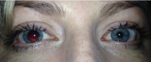
A 33-year-old woman with no past medical history presented to the emergency department with asymmetric pupils. At 7:30am while putting on makeup, she noted her pupils were equal in size. One hour later, she developed light sensitivity in her right eye, and soon after noticed her right pupil was significantly enlarged. She denied headache, facial or extremity weakness, dysarthria, or ataxia. On exam, her left pupil was reactive from 4 to 3 mm and her right pupil was sluggishly reactive and 8 mm (Figure). No abnormalities in her visual acuity, extraocular movement or fundoscopic exam were detected. Neurologic consultation was obtained, but the patient had an unremarkable brain computed tomography (CT)/CT-Angiography and magnetic resonance imaging.
A tonic pupil results from parasympathetic denervation at the level of the ciliary ganglion. It is characterized by a large, regular pupil with decreased response to light but preserved or enhanced constriction to accommodation, segmental iris constriction, vermiform movements of the pupillary border, and hypersensitivity to pharmacologic constricting agents.1–4 The diagnosis was established in consultation with ophthalmology and confirmed with rapid miotic response of the affected pupil to 0.125% pilocarpine drop.1,2 Most cases are idiopathic, occurring in women 20–40 years of age, and referred to as the Adie’s tonic pupil, though this disorder can be due to local disorders within the orbit, including tumor, inflammation, trauma, surgery, ischemia or infection. 3 Most patients do not require any treatment and can be reassured once the diagnosis is confirmed.
Footnotes
Supervising Section Editor: Sean O. Henderson, MD
Submission history: Submitted July 5, 2012; Accepted July 30, 2012
Full text available through open access at http://escholarship.org/uc/uciem_westjem
DOI: 10.5811/westjem.2012.7.12923
Address for Correspondence: Hemal K. Kanzaria, MD, Robert Wood Johnson Foundation Clinical Scholar®, University of California Los Angeles, Department of Emergency Medicine, Los Angeles, California
Email: Hemal.Kanzaria@gmail.com
Conflicts of Interest: By the WestJEM article submission agreement, all authors are required to disclose all affiliations, funding sources, and financial or management relationships that could be perceived as potential sources of bias. The authors disclosed none.
REFERENCES
1. Iannetti P, Spalice A, Iannetti L, et al. Residual and persistent Adie’s pupil after pediatric ophthalmoplegic migraine. Pediatric neurology. 2009;41:204–6. [PubMed]
2. Tafakhori A, Aghamollaii V, Modabbernia A, et al. Adie’s pupil during migraine attack: case report and review of literature. Acta neurologica Belgica. 2011;111:66–8. [PubMed]
3. Purvin VA. Adie’s tonic pupil secondary to migraine. Journal of neuroophthalmology : the official journal of the North American Neuro-Ophthalmology Society. 1995;15:43–4.
4. Moeller JJ, Maxner CE. The dilated pupil: an update. Current neurology and neuroscience reports. 2007;7:417–22. [PubMed]


