Treatment Protocol Assessment

Treatment Protocol Assessment

Blunt Trauma Abdominal and Pelvic Computed Tomography Has Low Yield for Injuries in More Than One Anatomic Region

Rodriguez, MD, et al.

In this retrospective study, we reviewed the imaging and hospital course of a consecutive sample of blunt trauma activation patients older than 14 years of age who received paired A/P CT during their blunt trauma assessments at an urban Level I trauma center from April through October 2014.

Read More
Treatment Protocol Assessment

Even the Thinnest Salami Contains Some Meat

Walsh, MB, et al.

This study is a secondary analysis of a subset of patients admitted to the hospital or pediatric intensive care unit in the Multicenter Airway Research Collaboration (http://www.emnet-usa.org).

Read More
Treatment Protocol Assessment

Head CT for Minor Head Injury Presenting to the Emergency Department in the Era of Choosing Wisely

DeAngelis, MD, et al.

The Choosing Wisely campaign currently recommends avoiding computed tomography (CT) of the head in low-risk emergency department (ED) patients with minor head injury, based on validated decision rules. The objective of this study was to evaluate adherence to the Choosing Wisely campaign’s recommendations regarding head CT imaging of patients with minor head injury in the ED.

Read More
Treatment Protocol Assessment

Emergency Department Pain Management Following Implementation of a Geriatric Hip Fracture Program

Scott D. Casey, MS, et al.

Over 300,000 patients in the United States sustain low-trauma fragility hip fractures annually. Multidisciplinary geriatric fracture programs (GFP) including early, multimodal pain management reduce morbidity and mortality. Our overall goal was to determine the effects of a GFP on the emergency department (ED) pain management of geriatric fragility hip fractures.

Read More
Treatment Protocol Assessment
Emergency Department Management of Suspected Calf-Vein Deep Venous Thrombosis: A Diagnostic Algorithm

Volume 17, Issue 4, July 2016
Levi Kitchen, MD et al.

Unilateral leg swelling with suspicion of deep venous thrombosis (DVT) is a common emergency department (ED) presentation. Proximal DVT (thrombus in the popliteal or femoral veins) can usually be diagnosed and treated at the initial ED encounter. When proximal DVT has been ruled out, isolated calf-vein deep venous thrombosis (IC-DVT) often remains a consideration. The current standard for the diagnosis of IC-DVT is whole-leg vascular duplex ultrasonography (WLUS), a test that is unavailable in many hospitals outside normal business hours. When WLUS is not available from the ED, recommendations for managing suspected IC-DVT vary. The objectives of the study is to use current evidence and recommendations to (1) propose a diagnostic algorithm for IC-DVT when definitive testing (WLUS) is unavailable; and (2) summarize the controversy surrounding IC-DVT treatment.

Read More
Treatment Protocol Assessment
Prospective Validation of Modified NEXUS Cervical Spine Injury Criteria in Low-risk Elderly Fall Patients

Volume 17, Issue 3, May 2016
John Tran, MD et al.

Introduction: The National Emergency X-radiography Utilization Study (NEXUS) criteria are used
extensively in emergency departments to rule out C-spine injuries (CSI) in the general population.
Although the NEXUS validation set included 2,943 elderly patients, multiple case reports and the
Canadian C-Spine Rules question the validity of applying NEXUS to geriatric populations. The
objective of this study was to validate a modified NEXUS criteria in a low-risk elderly fall population
with two changes: a modified definition for distracting injury and the definition of normal mentation.
Methods: This is a prospective, observational cohort study of geriatric fall patients who presented
to a Level I trauma center and were not triaged to the trauma bay. Providers enrolled nonintoxicated
patients at baseline mental status with no lateralizing neurologic deficits. They
recorded midline neck tenderness, signs of trauma, and presence of other distracting injury.
Results: We enrolled 800 patients. One patient fall event was excluded due to duplicate
enrollment, and four were lost to follow up, leaving 795 for analysis. Average age was 83.6 (range
65-101). The numbers in parenthesis after the negative predictive value represent confidence
interval. There were 11 (1.4%) cervical spine injuries. One hundred seventeen patients had midline
tenderness and seven of these had CSI; 366 patients had signs of trauma to the face/neck, and
10 of these patients had CSI. Using signs of trauma to the head/neck as the only distracting injury
and baseline mental status as normal alertness, the modified NEXUS criteria was 100% sensitive
(CI [67.9-100]) with a negative predictive value of 100 (98.7-100).
Conclusion: Our study suggests that a modified NEXUS criteria can be safely applied to lowrisk
elderly falls.

Read More
Treatment Protocol Assessment

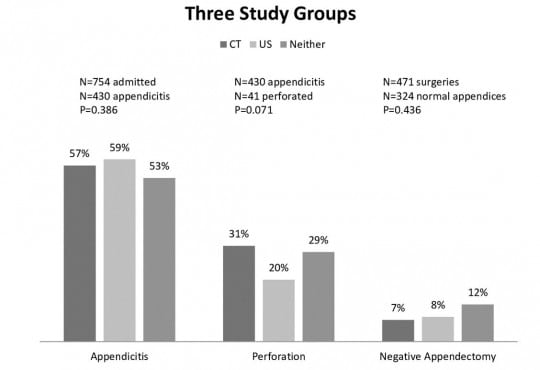
Abdominal CT Does Not Improve Outcome for Children with Suspected Acute Appendicitis

Volume 16, Issue 7, December 2015.
Danielle I. Miano, BS

Introduction: Acute appendicitis in children is a clinical diagnosis, which often requires preoperative
confirmation with either ultrasound (US) or computed tomography (CT) studies. CTs expose children to
radiation, which may increase the lifetime risk of developing malignancy. US in the pediatric population
with appropriate clinical follow up and serial exam may be an effective diagnostic modality for many
children without incurring the risk of radiation. The objective of the study was to compare the rate of
appendiceal rupture and negative appendectomies between children with and without abdominal CTs;
and to evaluate the same outcomes for children with and without USs to determine if there were any
associations between imaging modalities and outcomes.

Methods: We conducted a retrospective chart review including emergency department (ED) and inpatient
records from 1/1/2009–2/31/2010 and included patients with suspected acute appendicitis.

Results: 1,493 children, aged less than one year to 20 years, were identified in the ED with suspected
appendicitis. These patients presented with abdominal pain who had either a surgical consult or an
abdominal imaging study to evaluate for appendicitis, or were transferred from an outside hospital or
primary care physician office with the stated suspicion of acute appendicitis. Of these patients, 739 were
sent home following evaluation in the ED and did not return within the subsequent two weeks and were
therefore presumed not to have appendicitis. A total of 754 were admitted and form the study population,
of which 20% received a CT, 53% US, and 8% received both. Of these 57%, 95% CI [53.5,60.5] had
pathology-proven appendicitis. Appendicitis rates were similar for children with a CT (57%, 95% CI
[49.6,64.4]) compared to those without (57%, 95% CI [52.9,61.0]). Children with perforation were similar
between those with a CT (18%, 95% CI [12.3,23.7]) and those without (13%, 95% CI [10.3,15.7]).
The proportion of children with a negative appendectomy was similar in both groups: CT (7%, 95% CI
[2.1,11.9]), US (8%, 95% CI [4.7,11.3]) and neither (12%, 95% CI [5.9,18.1]).

Conclusion: In this uncontrolled study, the accuracy of preoperative diagnosis of appendicitis and
the incidence of pathology-proven perforation appendix were similar for children with suspected acute
appendicitis whether they had CT, US or neither imaging, in conjunction with surgical consult. The
imaging modality of CT was not associated with better outcomes for children presenting to the ED with
suspected appendicitis.

Read More

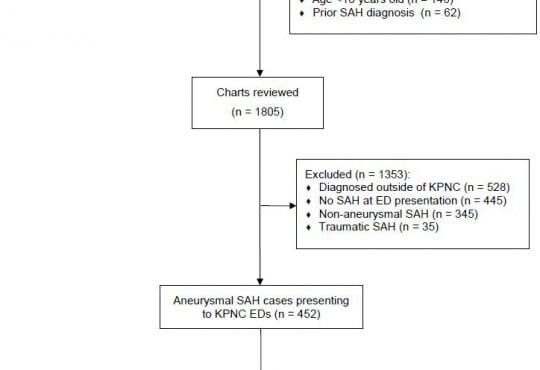
Sensitivity of a Clinical Decision Rule and Early Computed Tomography in Aneurysmal Subarachnoid Hemorrhage

Volume 16, Issue 5, September 2015.
Dustin G. Mark, MD, et al.

Introduction: Application of a clinical decision rule for subarachnoid hemorrhage, in combination with
cranial computed tomography (CT) performed within six hours of ictus (early cranial CT), may be able
to reasonably exclude a diagnosis of aneurysmal subarachnoid hemorrhage (aSAH). This study’s
objective was to examine the sensitivity of both early cranial CT and a previously validated clinical
decision rule among emergency department (ED) patients with aSAH and a normal mental status.
Methods: Patients were evaluated in the 21 EDs of an integrated health delivery system between
January 2007 and June 2013. We identified by chart review a retrospective cohort of patients
diagnosed with aSAH in the setting of a normal mental status and performance of early cranial CT.
Variables comprising the SAH clinical decision rule (age >40, presence of neck pain or stiffness,
headache onset with exertion, loss of consciousness at headache onset) were abstracted from the
chart and assessed for inter-rater reliability.
Results: One hundred fifty-five patients with aSAH met study inclusion criteria. The sensitivity of
early cranial CT was 95.5% (95% CI [90.9-98.2]). The sensitivity of the SAH clinical decision rule
was also 95.5% (95% CI [90.9-98.2]). Since all false negative cases for each diagnostic modality
were mutually independent, the combined use of both early cranial CT and the clinical decision rule
improved sensitivity to 100% (95% CI [97.6-100.0]).
Conclusion: Neither early cranial CT nor the SAH clinical decision rule demonstrated ideal
sensitivity for aSAH in this retrospective cohort. However, the combination of both strategies might
optimize sensitivity for this life-threatening disease.

Read More
Treatment Protocol Assessment
Triple Rule Out versus CT Angiogram Plus Stress Test for Evaluation of Chest Pain in the Emergency Department

Volume 16, Issue 5, September 2015.
Kelly N. Sawyer, MD, MS, et al.

Introduction: Undifferentiated chest pain in the emergency department (ED) is a diagnostic
challenge. One approach includes a dedicated chest computed tomography (CT) for pulmonary
embolism or dissection followed by a cardiac stress test (TRAD). An alternative strategy is a
coronary CT angiogram with concurrent chest CT (Triple Rule Out, TRO). The objective of this study
was to describe the ED patient course and short-term safety for these evaluation methods.
Methods: This was a retrospective observational study of adult patients presenting to a large,
community ED for acute chest pain who had non-diagnostic electrocardiograms (ECGs) and normal
biomarkers. We collected demographics, ED length of stay, hospital costs, and estimated radiation
exposures. We evaluated 30-day return visits for major adverse cardiac events.
Results: A total of 829 patients underwent TRAD, and 642 patients had TRO. Patients undergoing
TRO tended to be younger (mean 52.3 vs 56.5 years) and were more likely to be male (42.4% vs.
30.4%). TRO patients tended to have a shorter ED length of stay (mean 14.45 vs. 21.86 hours), to
incur less cost (median $449.83 vs. $1147.70), and to be exposed to less radiation (median 7.18 vs.
16.6mSv). No patient in either group had a related 30-day revisit.
Conclusion: Use of TRO is feasible for assessment of chest pain in the ED. Both TRAD and TRO
safely evaluated patients. Prospective studies investigating this diagnostic strategy are needed to
further assess this approach to ED chest pain evaluation.

Read More
Treatment Protocol Assessment
Comparing an Unstructured Risk Stratification to Published Guidelines in Acute Coronary Syndromes

Volume 16, Issue 5, September 2015.
Ann-Jean CC. Beck, MD

Introduction: Guidelines are designed to encompass the needs of the majority of patients with a
particular condition. The American Heart Association (AHA) in conjunction with the American College
of Cardiology (ACC) and the American College of Emergency Physicians (ACEP) developed risk
stratification guidelines to aid physicians with accurate and efficient diagnosis and management
of patients with acute coronary syndrome (ACS). While useful in a primary care setting, in the
unique environment of an emergency department (ED), the feasibility of incorporating guidelines
into clinical workflow remains in question. We aim to compare emergency physicians’ (EP) clinical
risk stratification ability to AHA/ACC/ACEP guidelines for ACS, and assessed each for accuracy in
predicting ACS.
Methods: We conducted a prospective observational cohort study in an urban teaching hospital
ED. All patients presenting to the ED with chest pain who were evaluated for ACS had two risk
stratification scores assigned: one by the treating physician based on clinical evaluation and
the other by the AHA/ACC/ACEP guideline aforementioned. The patient’s ACS risk stratification
classified by the EP was compared to AHA/ACC/ACEP guidelines. Patients were contacted at 30
days following the index ED visit to determine all cause mortality, unscheduled hospital/ED revisits,
and objective cardiac testing performed.
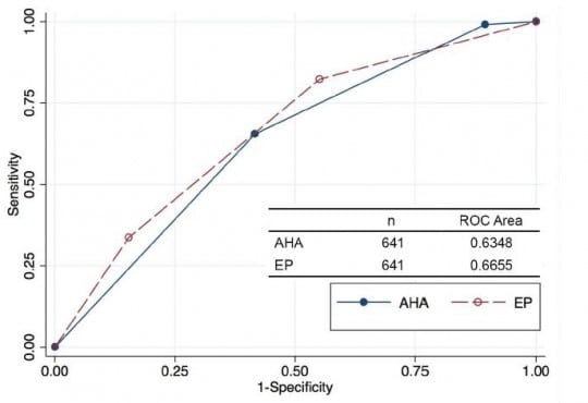
Results: We enrolled 641 patients presenting for evaluation by 21 different EPs. There was a
difference between the physician’s clinical assessment used in the ED, and the AHA/ACC/ACEP
task force guidelines. EPs were more likely to assess patients as low risk (40%), while AHA/ACC/
ACEP guidelines were more likely to classify patients as intermediate (45%) or high (45%) risk. Of
the 119 (19%) patients deemed high risk by EP evaluation, 38 (32%) were diagnosed with ACS.
AHA/ACC/ACEP guidelines classified only 57 (9%) patients low risk with 56 (98%) of those patients
diagnosed with no ACS.
Conclusion: In the ED, physicians are more efficient at correctly placing patients with underlying
ACS into a high-risk category. A small percentage of patients were considered low risk when
applying AHA/ACC/ACEP guidelines, which demonstrates how clinical insight is often required to
make an efficient assessment of cardiac risk and established criteria may be overly conservative
when applied to an acute care population.

Read More
Treatment Protocol Assessment
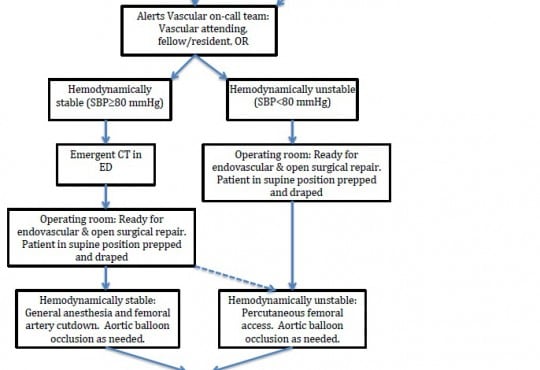
Case Series of Patients with Ruptured Abdominal Aortic Aneurysm

Volume 16, Issue 3, May 2015
Taylor Spencer, MD, MPH, et al.

Traditionally, patients with suspected ruptured abdominal aortic aneurysm (rAAA) are taken immediately for operative repair. Computed tomography (CT) has been considered contraindicated. However, with the emergence of endovascular repair, this approach to suspected rAAA could be changing.

Read More
Treatment Protocol Assessment
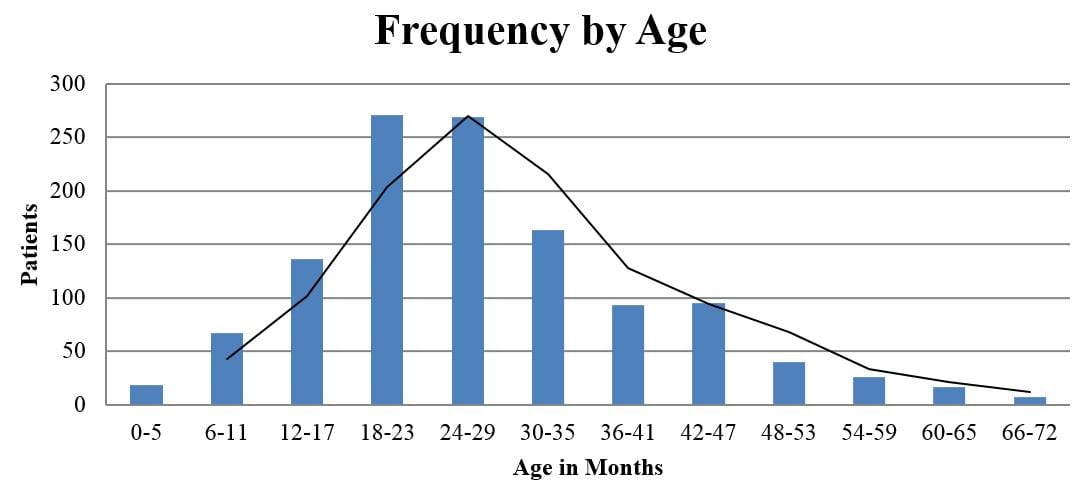
Epidemiology of Nursemaid’s Elbow

Volume 15, Issue 4, July 2014
Sarah Vitello, DO et al.

To provide an epidemiological description of radial head subluxation, also known as nursemaid’s elbow, from a database of emergency department visits.

Read More
Treatment Protocol Assessment

Predictors of Unattempted Central Venous Catheterization in Septic Patients Eligible for Early Goal-directed Therapy

Volume XV, Issue 1, February 2014
David R. Vinson, MD et al.

We sought to determine the association of relative normotension (sustained systolic blood pressure >90 mmHg independent of or in response to an initial crystalloid resuscitation of 20 mL/kg), obesity (body mass index [BMI] ≥30), moderate thrombocytopenia (platelet count <50,000 per μL), and coagulopathy (international normalized ratio ≥2.0) with unattempted CVC in EGDT-eligible patients.

Read More

Contact Information

WestJEM/ Department of Emergency Medicine
UC Irvine Health

3800 W Chapman Ave Ste 3200
Orange, CA 92868, USA
Phone: 1-714-456-6389
Email: editor@westjem.org

CC-BY_icon.svg

WestJEM
ISSN: 1936-900X
e-ISSN: 1936-9018

CPC-EM
ISSN: 2474-252X

Our Philosophy

Emergency Medicine is a specialty which closely reflects societal challenges and consequences of public policy decisions. The emergency department specifically deals with social injustice, health and economic disparities, violence, substance abuse, and disaster preparedness and response. This journal focuses on how emergency care affects the health of the community and population, and conversely, how these societal challenges affect the composition of the patient population who seek care in the emergency department. The development of better systems to provide emergency care, including technology solutions, is critical to enhancing population health.