
| Author | Affiliation |
|---|---|
| Peter Moffett, MD | Department of Emergency Medicine, Carl R. Darnall Army Medical Center, Fort Hood, Texas |
An approximately 30 year-old Ugandan male was found unresponsive on an American base in Iraq. The patient was altered and combative with no signs of trauma. A chest x-ray was performed showing an enlarged cardiac silhouette and pulmonary edema. The patient required intubation during which a large amount of edema fluid was produced. A computed tomography (CT) (Figure) showed severe calcifications of both the aortic and mitral valves.
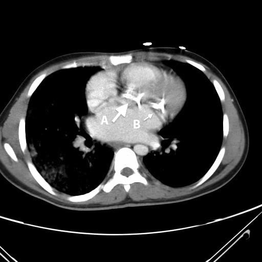
Intravenous contrast enhanced computed tomography of the chest showing severe calcifications (arrows) of both aortic (A) and mitral (B) valves.
Shortly after CT the patient went into cardiac arrest. The patient had a bedside echocardiogram showing severe bi-valvular heart failure and died shortly thereafter. The diagnosis of rheumatic heart disease with severe cardiomyopathy was considered the most likely cause of his presentation.
Rheumatic heart disease is a major cause of cardiovascular death in developing countries and affects between 15 and 20 million people worldwide.1 The disease is a sequella of one or more episodes of acute rheumatic fever with subsequent valvular calcification and damage. The pathophysiology is felt to be an aberrant immune response to certain strains of Streptococcus pyogenes infection. The mitral valve is most commonly affected, but aortic valve involvement is also seen as the disease progresses. Patients may not recall any preceding events and present with shortness of breath between ages 20 and 50.1 Diagnosis of rheumatic disease is often made by echocardiogram when the diagnosis is suspected. Without intervention, the disease progresses to severe valvular disease with heart failure.
Footnotes
Address for Correspondence: Peter Moffett, MD. Carl R Darnall Army Medical Center, 36000 Darnall Loop, ER Box 31, Fort Hood, TX 76544. Email: pmoff2@gmail.com. 11 / 2013; 14:640 – 640
Submission history: Revision received June 2, 2013; Submitted June 17, 2013; Accepted July 15, 2013
Conflicts of Interest: By the WestJEM article submission agreement, all authors are required to disclose all affiliations, funding sources and financial or management relationships that could be perceived as potential sources of bias. The authors disclosed none. The opinions or assertions contained herein are the private views of the author and are not to be construed as official or as reflecting the views of the United States Army, Department of Defense, or the United States government.
REFERENCES
1. Marijon E, Mirabel M, Celermajer DS, Jouven X Rheumatic heart disease. Lancet. 2012; 379:953-964