
| Author | Affiliation |
|---|---|
| Jesse Wray, MD | San Antonio Uniformed Services Health Education Consortium, Department of Emergency Medicine, Fort Sam Houston, Texas |
| Rachel E. Bridwell, MD | San Antonio Uniformed Services Health Education Consortium, Department of Emergency Medicine, Fort Sam Houston, Texas |
| Michael J. Yoo, MD | San Antonio Uniformed Services Health Education Consortium, Department of Emergency Medicine, Fort Sam Houston, Texas |
| Christopher N. Belcher, MD | San Antonio Uniformed Services Health Education Consortium, Department of Emergency Medicine, Fort Sam Houston, Texas |
| Joshua J. Oliver, MD | San Antonio Uniformed Services Health Education Consortium, Department of Emergency Medicine, Fort Sam Houston, Texas |
ABSTRACT
Case Presentation
We describe a case of spontaneous partial segmental thrombosis of the corpus cavernosum (PSTCC).
Discussion
PSTCC is a rare condition in which thrombus formation occurs in the corpus cavernosum. This condition is managed in conjunction with a urologist, and management typically includes anticoagulation and pain control.
CASE PRESENTATION
A 39-year-old man presented to the emergency department for two days of worsening pain and swelling to the base of his penis. The patient denied trauma or a history of coagulopathy, had a non-contributory sexual history, and no recent use of erectile dysfunction medications. Examination demonstrated mild swelling to the penile base without evidence of hernia, infection, or shaft injury.
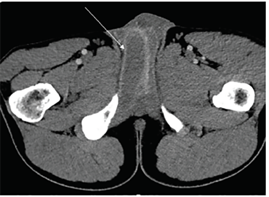
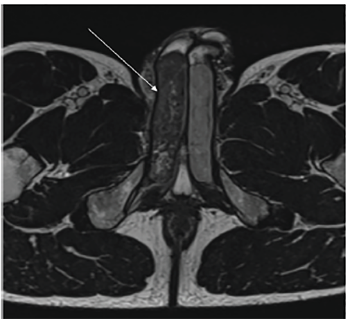
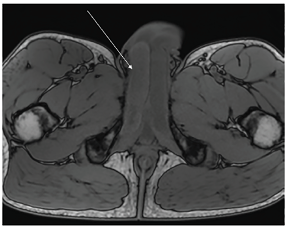
Computed tomography (CT) revealed penile asymmetry (Image 1). Ultrasound demonstrated asymmetric fullness of the right corpus cavernosum. Pelvis magnetic resonance imaging (MRI) revealed an enlarged appearance of the right corpus cavernosum with hypointense T2 signal (Image 2) and hyperintense T1 signal (Image 3). These findings were consistent with a partial segmental thrombosis of the right corpus cavernosum (PSTCC). The patient was admitted for pain control and discharged after symptom resolution with anticoagulation therapy. Upon outpatient follow-up, the patient had no persistent complications.
DISCUSSION
PSTCC is a rare condition that manifests as penile or perineal pain and swelling. Thrombus formation likely arises secondary to microtrauma, thrombophilia, hemoglobinopathies and, rarely, medication side effect.1,2 Ultrasonography or MRI are recommended diagnostic modalities, while CT is reportedly suboptimal due to decreased sensitivity for this condition.3 Our case departs from the literature as CT and MRI were most useful. Additionally, because CT clearly demonstrates the pathology in this case, it may be a better diagnostic modality than previously reported in this rare phenomenon and serve as a rapid diagnostic tool in some cases. Early urologic consultation is recommended, with typical management consisting of anticoagulation and pain control.2 PSTCC has an overall favorable prognosis rarely incurring long-term complications.3
CPC-EM Capsule
What do we already know about this clinical entity?
Partial segmental thrombosis of the corpus cavernosum (PSTCC) is a rare condition classically diagnosed with ultrasound or magnetic resonance imaging.
What is the major impact of the image(s)?
Although computed tomography (CT) has not been previously recommended for identifying this pathology, our case demonstrates that PSTCC can be clearly identified with CT.
How might this improve emergency medicine practice?
This example of a rare pathology that may go unrecognized by emergency providers demonstrates the use of CT to aid in diagnosis.
Footnotes
Section Editor: Rick A. McPheeters, DO
Full text available through open access at http://escholarship.org/uc/uciem_cpcem
The authors attest that their institution requires neither Institutional Review Board approval, nor patient consent for publication of this image in emergency medicine. Documentation on file.
Address for Correspondence: Jesse Wray, MD, San Antonio Uniformed Services Health Education Consortium, Department of Emergency Medicine, 3551 Roger Brooke Dr, Fort Sam Houston, TX 78234. Email: jesse.p.wray.mil@mail.mil. 4:497 – 498
Submission history: Revision received April 26, 2020; Submitted June 15, 2020; Accepted July 3, 2020
Conflicts of Interest: By the CPC-EM article submission agreement, all authors are required to disclose all affiliations, funding sources and financial or management relationships that could be perceived as potential sources of bias. The view(s) expressed herein are those of the author(s) and do not reflect the official policy or position of Brooke Army Medical Center, the U.S. Army Medical Department, the U.S. Army Office of the Surgeon General, the Department of the Army, the Department of the Air Force and Department of Defense or the U.S. Government. The authors disclosed none.
REFERENCES
1. Christodoulidou M, Parnham A, Ramachandran N, et al. Partial segmental thrombosis of the corpus cavernosum presenting with perineal pain. BMJ Case Rep. 2016;2016.
2. Hoyerup PH, Azawi NH. Partial priapism. BMJ Case Reports. 2013;2013.
3. Smetana A, Driver B, Gajic S, et al. Partial segmental thrombosis of the corpus cavernosum presenting to the ED. Am J Emerg Med. 2016;34(6):1182.e3-5.