
| Author | Affiliation |
|---|---|
| Caleb Patrick Canders, MD | David Geffen School of Medicine, University of California Los Angeles, Department of Emergency Medicine, Los Angeles, California |
| Lynne McCullough, MD | David Geffen School of Medicine, University of California Los Angeles, Department of Emergency Medicine, Los Angeles, California |
CASE
A 67-year-old man presented with painful oral and skin lesions developing over the last 2 months. The lesions initially formed in his mouth and lips, and slowly spread to his torso, groin, and extremities (Figure). The lesions began as blisters that broke easily and were exquisitely painful to light touch. In the past few days, he also developed painful lesions on his eyes that were associated with redness and photophobia. The patient saw his primary care doctor at the onset of illness and failed to improve with courses of azithromycin, ciprofloxacin, and tetracycline. He was no longer able to tolerate anything by mouth because of pain and he had lost a significant amount of weight. His skin exam was notable for Nikolsky’s sign.
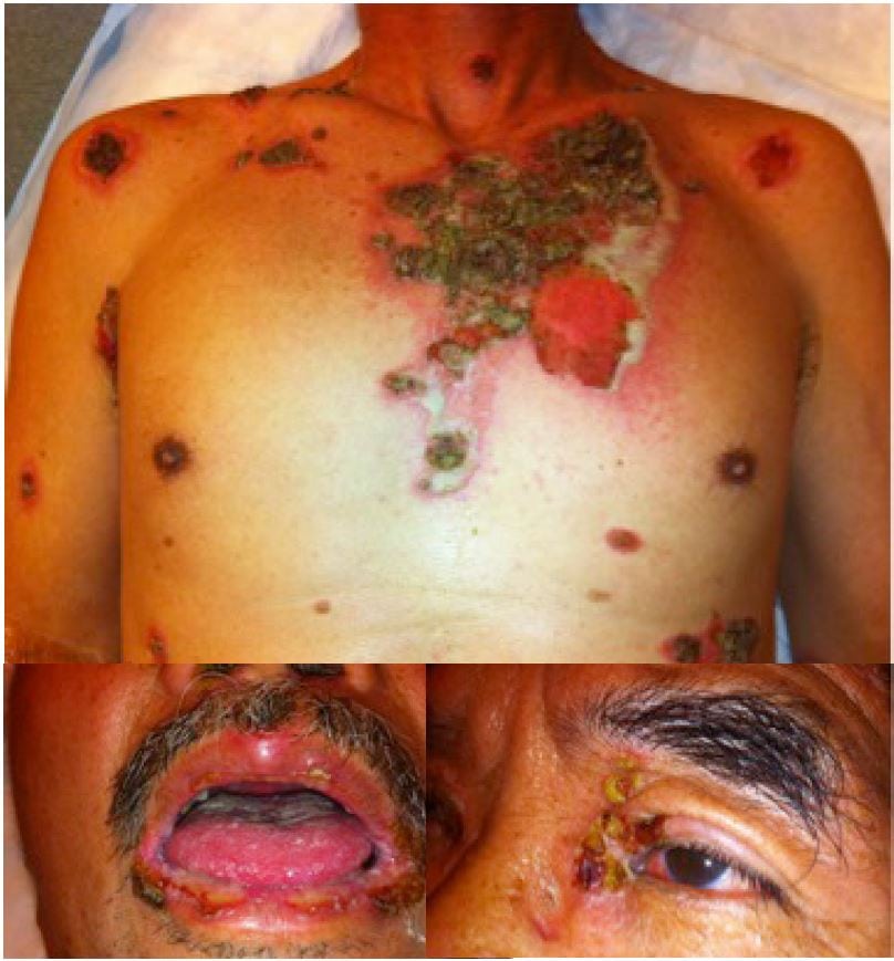
Extensive erosions and flaccid bullae associated with autoimmune blistering disorder.
DIAGNOSIS
Pemphigus vulgaris is an autoimmune blistering disorder mediated by auto-antibodies against epidermal cell antigens.1 It has an incidence of approximately 1 in 100,000, and the usual age of onset is 40–60 years of age.2 Patients present with painful, flaccid bullae that rupture and form erosions (Figure). Shearing stress on skin can lead to development of new erosions (Nikolsky’s sign). Oral lesions are the initial symptom in 50–60% patients and may precede cutaneous lesions by months. Diagnosis is confirmed by histology and direct immunofluorescence of peri-lesional skin. Mortality is 70% if untreated, and primarily results from sepsis, fluid loss and malnutrition from oral lesions.3
Our patient was admitted for fluid resuscitation and intravenous steroids, and had a skin biopsy consistent with pemphigus vulgaris.
Footnotes
Address for Correspondence: Caleb Patrick Canders, MD. Department of Emergency Medicine, David Geffen School of Medicine at UCLA, 924 Westwood Boulevard, Suite 300 | Box 951777, Los Angeles, CA 90095-1777. Email: caleb.canders@gmail.com. 11 / 2013; 14:645 – 645
Submission history: Revision received July 1, 2013; Accepted July 15, 2013
Conflicts of Interest: By the WestJEM article submission agreement, all authors are required to disclose all affiliations, funding sources and financial or management relationships that could be perceived as potential sources of bias. The authors disclosed none.
REFERENCES
1. Zaidi Z, Lanigan SW Bullous disorders. Dermatology in Clinical Practice. 2010; :233-252
2. Greenberg MI, Rubenstein B Diagnosis: pemphigus vulgaris. Emergency Medicine News. 2002; 24:14
3. Sperber BR, Martin LK, Murrell DF Pemphigus. Evidence-based Dermatology. 2008; :581-594