
| Author | Affiliation |
|---|---|
| Amanda H. Westfall, MD | Baylor University Medical Center, Department of Emergency Medicine, Dallas, Texas |
| John S. Garrett, MD | Baylor University Medical Center, Department of Emergency Medicine, Dallas, Texas |
ABSTRACT
Emergency physicians often rely on a “triple-rule-out” computed tomography (CT) where image acquisition is timed to obtain image quality equivalent to dedicated coronary CT angiography, pulmonary CT angiography, and thoracic aorta CT angiography. This case highlights the importance of obtaining CT angiography dedicated to the aorta in the setting of high clinical suspicion for aortic disease if initial CT pulmonary angiogram is negative for aortic disease.
CASE PRESENTATION
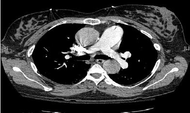
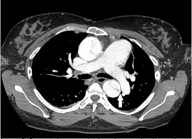
A 39-year-old woman with twin gestation at 25 weeks presented to the emergency department with syncope. Her past medical history was significant for Marfan’s syndrome. Physical exam demonstrated confusion, hypotension, and a normal cardiopulmonary exam with equal pulses in all extremities. The patient denied chest pain. Differential diagnosis included aortic dissection and, given her pregnant state, pulmonary embolism. To minimize radiation exposure, a computed tomography (CT) pulmonary angiogram (PA) was obtained to evaluate for pulmonary embolism and aortic dissection (Image 1). Due to a negative CTPA and high suspicion for aortic disease, CT of the thoracic aorta was obtained (Image 2).
DISCUSSION
Type A Aortic Dissection
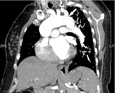
CT of the thoracic aorta revealed a dissection from the aortic root to the abdominal aorta and involving the left common carotid (Image 3). The patient was sent to the operating room for fenestration of the thoracoabdominal aorta and subsequent replacement of the ascending aorta and hemiarch. Her twins were delivered by emergent cesarean section at 28 weeks gestation. Mother was discharged to a rehabilitation facility and then home.
Thoracic CT imaging may evaluate for pulmonary embolism, aortic disease, and coronary artery disease. This can be accomplished through a “triple-rule-out” CT, where image acquisition is timed to obtain image quality equivalent to dedicated coronary, pulmonary and thoracic aorta CT angiography with high sensitivity and specificity.1 Emergency physicians should be aware of the significant limitations related to the contrast bolus timing, especially if a CTPA is used to screen for aortic pathology. If intravenous contrast is not within the aorta at the time of image acquisition, false negative results can occur, making aortic dissection invisible.2
CPC-EM Capsule
What do we already know about this clinical entity?
Type A Aortic dissection is a life-threatening disease with high morbidity and mortality. It requires early diagnosis, most often accomplished via computed tomography (CT) imaging.
What is the major impact of the image(s)?
These comparison images demonstrate the importance of obtaining CT angiography dedicated to the aorta, even if initial CT pulmonary angiogram is negative for aortic disease.
How might this improve emergency medicine practice?
These comparison images demonstrate the importance of obtaining CT angiography dedicated to the aorta, even if initial CT pulmonary angiogram is negative for aortic disease.
Footnotes
Section Editor: Steven Walsh, MD
Full text available through open access at http://escholarship.org/uc/uciem_cpcem
Documented patient informed consent and/or Institutional Review Board approval has been obtained and filed for publication of this case report.
Address for Correspondence: Amanda H. Westfall, MD, Baylor University Medical Center, Department of Emergency Medicine, 3500 Gaston Avenue, Dallas, TX 75214. Email: amanda.westfall@bswhealth.org. 3:316 – 317
Submission history: Revision received January 26, 2019; Submitted May 21, 2019; Accepted May 28, 2019
Conflicts of Interest: By the CPC-EM article submission agreement, all authors are required to disclose all affiliations, funding sources and financial or management relationships that could be perceived as potential sources of bias. The authors disclosed none.
REFERENCES
1. McMahon MA, Squirrell CA. Multidetector CT of the aortic dissection: a pictorial review. Radiographics. 2010;30(2):445-60.
2. Dähnert W. Cardiovascular disorders: aortic dissection. Radiology Review Manual. 2003:607-9.