
| Author | Affiliation |
|---|---|
| Jeremy N. Johnson, DO | Madigan Army Medical Center/University of Washington Emergency Medicine Residency, Fort Lewis, WA |
| Daniel F. McBride, MD, MPH | Madigan Army Medical Center/University of Washington Emergency Medicine Residency, Fort Lewis, WA |
| Steve Crandall, MD | Madigan Army Medical Center/University of Washington Emergency Medicine Residency, Fort Lewis, WA |
| Christopher Kang, MD | Madigan Army Medical Center/University of Washington Emergency Medicine Residency, Fort Lewis, WA |
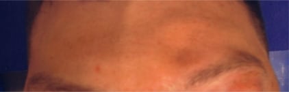
A 21-year-old man walked into the ED complaining of mild headache after colliding with another player during a baseball game. The patient denied loss of consciousness and noted numbness over his left forehead. He denied vision change, fluid from his nose or ears, dizziness or other injury. He had normal mentation, no neck pain, and an otherwise non-focal exam. He declined pain medication.
A visible depression is noted overlying the left frontal bone. (Figure 1.) An ultrasound (US) at the bedside demonstrates an isolated comminuted fracture of the left frontal sinus with depression of one segment into the sinus and an overlying flap created by the other fragment. The CT image confirmed this (Figure 2).

The force required to fracture the frontal sinus ranges from 800–2200 lbs, more than enough to cause associated injuries.1 Complications include: CSF leak, osteomyelitis, meningitis, brain abscess or mucopyocele, and cavernous sinus thrombosis. Operative repair is best performed by an ENT specialist using endoscopic techniques to assure good cosmetic outcome.2
Currently CT, which is the criterion reference for diagnosis of skull fractures, is most important to evaluate for inner table fractures or co-existing mid-face fractures.3 Despite the superiority of CT, US can be used in remote or austere environments. Research comparing CT to US for superficial facial fractures needs to be done to determine test characteristics.
Footnotes
The opinions and assertions contained herein are the private views of the authors and should not be construed as official or as reflecting the views of the Department of the Army or the Department of Defense.
Supervising Section Editor: J. Christian Fox, MD
Submission history: Submitted October 13, 2007; Revision Received January 25, 2009; Accepted January 26, 2009
Full text available through open access at http://escholarship.org/uc/uciem_westjem
Address for Correspondence: Jeremy N. Johnson, DO, Department of Emergency Medicine, Madigan Army Medical Center, Fort Lewis, WA
Email: jeremy.n.johnson@amedd.army.mil
Conflicts of Interest: By the WestJEM article submission agreement, all authors are required to disclose all affiliations, funding sources, and financial or management relationships that could be perceived as potential sources of bias. The authors disclosed none.
REFERENCES
1. Nahum AM. The biomechanics of maxillofacial trauma. Clin Plast Surg. 1975;2:59–64. [PubMed]
2. Strong EB, Kellman RM. Endoscopic repair of anterior table–frontal sinus fractures. Facial Plast Surg Clin North Am. 2006;14:25–9. [PubMed]
3. Sun JK, Lemay DR. Imaging of Facial Trauma. Neuroimaging Clin N Am. 2002;12:295–309.[PubMed]