
| Author | Affiliation |
|---|---|
| Daniel Thomas Naylor, MD | University of California, Department of Emergency Medicine, Davis, CA |
| Kapil R. Dhingra, MD, MBA | University of California, Department of Emergency Medicine, Davis, CA |
| Emily R. Andrada, MD | University of California, Department of Emergency Medicine, Davis, CA |
A two-year-old male presented to the emergency department (ED) with a four-day history of evening tactile fevers, measured to 38.1ºC at home, associated with a worsening cough, congestion, mild diarrhea, emesis, decreased oral intake and level of activity. His older brother arrived complaining of the same symptoms, and an older sister had also experienced an episodes of fever and emesis. The mother had not noticed any tugging at ears, shortness of breath, or evidence of abdominal pain. The patient’s fevers at home were controlled with acetaminophen. His vaccinations were up to date; he had no past medical history and his birth history was unremarkable. He had no known allergies.
On presentation to the ED, his vital signs were temperature of 37.6°C, heart rate of 144 beats per minute, blood pressure of 147/82 mmHg (kicking and screaming during measurement), respiratory rate of 28 breaths per minute, and oxygen saturation of 97%. The patient appeared to be in no acute distress, and did not appear toxic. He was interactive and smiling, and appropriately fussy during examination. He appeared to be breathing normally. Examination was significant for mildly decreased breath sounds over the right posterior thorax with no rales, wheezes, or rhonchi, and a normal respiratory rate with no accessory muscle use or retractions. He had normal heart sounds and a benign abdominal examination. He exhibited no pallor or cyanosis.
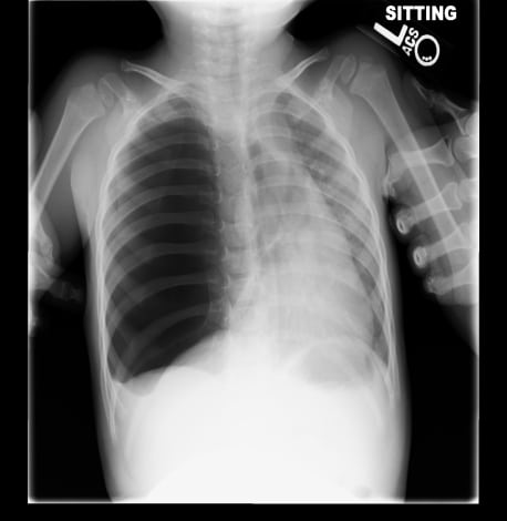
A chest x-ray was obtained (Figure 1), which revealed a large right-sided pneumothorax with a leftward shift of the mediastinum. A chest tube was inserted emergently and the patient was admitted to the hospital. Viral nasal swab culture was positive for parainfluenza virus. There was a persistent air leak in the chest tube. After an unsuccessful video-assisted thoracic surgery (VATS) pleurodesis, a thoracotomy was performed where evidence was found of a right lower lobe pedunculated lung cyst.
Footnotes
Supervising Section Editor: Sean Henderson, MD
Submission history: Submitted February 4, 2010; Revision Received February 7, 2010; Accepted February 22, 2010
Full text available through open access at http://escholarship.org/uc/uciem_westjem
Address for Correspondence: Daniel Naylor, MD, UC Davis Medical Center, Department of Emergency Medicine, 4150 V Street, PSSB Suite 2100, Sacramento, CA 95817
Email: daniel.naylor@ucdmc.ucdavis.edu
Conflicts of Interest: By the WestJEM article submission agreement, all authors are required to disclose all affiliations, funding sources, and financial or management relationships that could be perceived as potential sources of bias. The authors disclosed none.