
| Author | Affiliation |
|---|---|
| Tanner George Greiving, MD | Brooke Army Medical Center, Department of Emergency Medicine, San Antonio, Texas |
| Sumeru G. Mehta, MD | Methodist Hospital, Emergency Department, San Antonio, Texas |
ABSTRACT
Case Presentation
We describe a case of epipericardial fat necrosis.
Discussion
Epipericardial fat necrosis is an inflammatory condition in which the pericardial fat pad necrotizes resulting in surrounding inflammation. This condition mimics more ominous pathology in clinical presentation and radiographic findings. Management is supportive with oral analgesics.
CASE PRESENTATION
A 39-year-old male presented to the emergency department (ED) for three days of right-sided, pleuritic chest pain. The patient denied any preceding trauma or illness. Examination revealed no overlying skin changes or reproducible chest wall tenderness, although lung sounds were noted to be diminished near the right lung base. His vital signs were as follows: temperature of 98.2° Fahrenheit; respiratory rate of 17 breaths per minute; pulse oximetry of 95% on room air; blood pressure of 135/82 millimeters of mercury; and heart rate of 92 beats per minute.
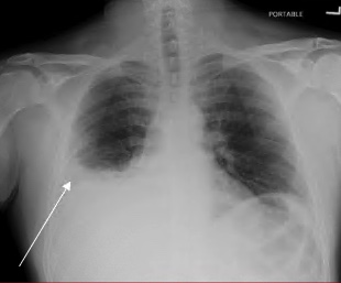
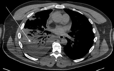
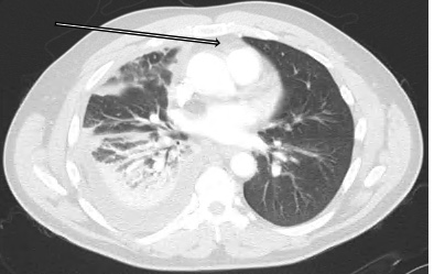
Chest radiograph revealed a right pleural effusion with right base consolidation suspicious for pneumonia (Image 1). Based on historical factors not consistent with pneumonia and discussion with the radiologist, a computed tomography (CT) chest without contrast was initially ordered. The CT chest demonstrated multilobular consolidations within the right lung with an associated moderate volume pleural effusion (Image 2). Subsequent concerns about possible pulmonary infarction as a cause of the pleural effusion prompted a CT angiogram. Computed tomography angiography demonstrated acute epipericardial fat necrosis with sympathetic right pleural effusion and right lung atelectasis (Image 3). The patient’s pain was controlled with oral analgesics during evaluation in the ED; he was then discharged home with continued oral analgesic therapy.
DISCUSSION
Epipericardial fat necrosis is a rare benign condition1 that presents as acute pleuritic chest pain. The description of symptoms may reflect that of more ominous pathologies including acute myocardial infarction, pulmonary embolism, or acute pericarditis.2 Epipericardial fat necrosis is characterized as a self-limited inflammatory process occurring inside the epipericardial fat—the tissue connecting the pericardial layer to the anterior thoracic wall.3 Findings on chest radiograph are typically non-specific.4 Computed tomography is the imaging modality of choice for diagnosis, although a CT angiogram may be warranted to rule out pulmonary embolism.5 Current management is supportive centering around oral analgesia, typically non-steroidal anti-inflammatories.3 A follow-up, non-contrast enhanced CT should be considered at 4–8 weeks to confirm expected healing.5
CPC-EM Capsule
What do we already know about this clinical entity?
Epipercardial fat necrosis is a self-limited, inflammatory condition which often causes chest pain and radiographic findings suggestive of more ominous pathologies.
What is the major impact of the image(s)?
These images demonstrate the characteristic fat pad changes in combination with radiographic findings that may also be present with more ominous pathologies.
How might this improve emergency medicine practice?
Early recognition of this etiology may reduce excessive imaging and aid in the initiation of appropriate management.
Footnotes
Section Editor: Manish Amin, DO
Full text available through open access at http://escholarship.org/uc/uciem_cpcem
Documented patient informed consent and/or Institutional Review Board approval has been obtained and filed for publication of this case report.
Address for Correspondence: Tanner G. Greiving, MD, Brooke Army Medical Center, Emergency Department, San Antonio Uniformed Services Health Education Consortium, Department of Emergency Medicine, 3551 Roger Brooke Dr, Fort Sam Houston, TX 78234. Email: tanner.greiving@gmail.com. 7:49 – 50
Submission history: Revision received June 29, 2022; Submitted October 16, 2022; Accepted October 20, 2022
Conflicts of Interest: By the CPC-EM article submission agreement, all authors are required to disclose all affiliations, funding sources and financial or management relationships that could be perceived as potential sources of bias. This review does not reflect the views or opinions of the U.S. government, Department of Defense, Defense Health Agency, U.S. Army, U.S. Air Force, or SAUSHEC EM Residency Program. The authors disclosed none.
REFERENCES
1. Hernandez D, Galimany J, Pernas JC, et al. Case 170: Pericardial fat necrosis. Radiology. 2011;259(3):919-22.
2. Pineda V, Cáceres J, Andreu J, et al. Epipericardial fat necrosis: radiologic diagnosis and follow-up. Am J Roentgenol. 2005;185(5):1234-6.
3. Giassi KdS, Costa AN, Bachion GH, et al. Epipericardial fat necrosis: an underdiagnosed condition. Br J Radiol. 2014;87(1038):20140118.
4. Jackson RC, Clagett OT, Mcdonald JR. Pericardial fat necrosis: report of three cases. J Thorac Surg. 1957;33(6):723-9.
5. Ataya D, Chowdhry AA, Mohammed TH. Epipericardial fat pad necrosis. J Thorac Imaging. 2011;26(4):W140-2.