
| Author | Affiliation |
|---|---|
| Fabrizio Elia, MD | High Dependency Unit, San Giovanni Bosco Hospital, Torino, Italy |
| Fiammetta Pagnozzi, MD | High Dependency Unit, San Giovanni Bosco Hospital, Torino, Italy |
| Paolo Busolli, MD | Radiology, San Giovanni Bosco Hospital, Torino, Italy |
| Franco Aprà, MD | High Dependency Unit, San Giovanni Bosco Hospital, Torino, Italy |
ABSTRACT
Volvulus is a frequent condition in patients presenting in the emergency department (ED) with abdominal pain. While cecal volvulus occurs more often in young patients, sigmoid volvulus is more common in elderly patients.
A 76-year-old man presented with abdominal pain and constipation. A previous ischemic stroke and subsequent neurological complications had left him bedridden for six years. Surgical history included tracheotomy and gastrostomy tube. On admission he was normotensive and afebrile but mildly tachycardic. Abdominal examination revealed severe distension and tenderness in the left lower quadrant with localized peritonitis. Laboratory tests yielded leukocytosis. Abdominal radiographs and computed tomography (CT) revealed a markedly dilated sigmoid colon in the presence of distal volvulus. (Figures 1 and and2)2) With clinical and laboratory signs suggestive of gangrene, the patient underwent open laparotomy. Sigmoid volvulus with necrotic bowel was detected; reduction of the volvulus and sigmoid resection was performed. No surgical complication occurred, but he ultimately succumbed to pulmonary complications.
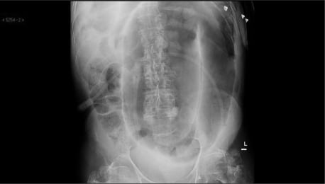
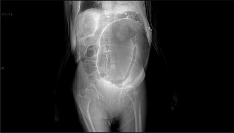
Volvulus is a common condition in patients presenting with abdominal pain. Cecal volvulus occurs more often in young patients, while sigmoid volvulus is more common in the elderly. Being bedridden, neuropsychiatric conditions and chronic constipation are common risk factors for sigmoid bowel involvement. Typical radiographs and CT findings include distended sigmoid loops with an inverted “U” shape and/or coffee-bean appearance (Figure 1 and and2).2). However, classic appearance is absent in one fourth of CT scans.2 Treatment requires detorsion of sigmoid loops. Endoscopic reduction is effective in most cases; however, recurrence is common. Surgical approach is indicated in suspected gangrene, perforation and for patients with recurrent volvulus.
The increasing access of elderly and frail patients in emergency departments entails an increase in diagnosis of sigmoid volvulus, raising clinical and ethical questions about appropriate treatment.3
Footnotes
Supervising Section Editor: Rick A. McPheeters, DO
Submission history: Submitted March 25, 2010; Revision Received April 28, 2010; Accepted May 6, 2010
Full text available through open access at http://escholarship.org/uc/uciem_westjem
Address for Correspondence: Fabrizio Elia, MD, Medicina d’Urgenza, Ospedale San Giovanni Bosco, Piazza Donatore del Sangue 3, 10154 Torino Italy
Email: fabrizioelia@yahoo.it
Conflicts of Interest: By the WestJEM article submission agreement, all authors are required to disclose all affiliations, funding sources, and financial or management relationships that could be perceived as potential sources of bias. The authors disclosed none.
REFERENCES
1. Tiah L, Goh SH. Sigmoid volvulus: diagnostic twists and turns. Eur J Emerg Med. 2006;13:84–7.[PubMed]
2. Levsky JM, Den EI, DuBrow RA, et al. CT findings of sigmoid volvulus. AJR Am J Roentgenol.2010;194:136–43. [PubMed]
3. Albaba M, Takahashi PY. 83-year-old woman with abdominal distention and constipation. Mayo Clin Proc. 2009;84:1126–9. [PMC free article] [PubMed]