| Author | Affiliation |
|---|---|
| Antonio Siniscalchi, MD | Department of Neurology, “Annunziata” Hospital, Cosenza, Italy Department of Neuroradiology, “Annunziata” Hospital, Cosenza, Italy |
| Luca Gallelli, MD, PhD | Department of Neurology, “Annunziata” Hospital, Cosenza, Italy Department of Experimental and Clinical Medicine, University Magna Grecia of Catanzaro, Regional Pharmacovigilance Center, Mater Domini University Hospital, Catanzaro, Italy |
| Olindo Di Benedetto, MD | Department of Neuroradiology, “Annunziata” Hospital, Cosenza, Italy |
| Giovambattista De Sarro, MD | Department of Experimental and Clinical Medicine, University Magna Grecia of Catanzaro, Regional Pharmacovigilance Center, Mater Domini University Hospital, Catanzaro, Italy |
ABSTRACT
Asterixis is not yet considered a common neurological sign of cerebellum infarction, and the pathogenic mechanism for asterixis remains elusive. We report a 58-year-old male with moderate hypertension who presented to our emergency department for acute headache in both cervical and occipital regions of the left side. About 2 hours later the patient developed ipsilateral asterixis in the upper left limb; 3 days later the asterixis disappeared. Magnetic resonance imaging of the brain disclosed cerebellar infarctions at the left superior cerebellar artery. In conclusion, we observed that a transitory asterixis associated with ipsilateral headache can be an initial clinical manifestation of ipsilateral cerebellar infarctions in the superior cerebellar artery area.
INTRODUCTION
Involuntary movements, such as chorea, dystonia, asterixis, and tremor, may occur as a consequence of stroke due to the involvement of basal ganglia or thalamus/ subthalamus.1,2 Involuntary movements caused by the anterior cerebral artery infarction have been reported.3 While asterixis has been described in 2 patients with ipsilateral cerebellar infarction, it is not yet considered a neurological sign of cerebellum infarction.2 We report a patient with unilateral headache and transitory asterixis of the upper left limb due to an acute cerebellar infarction in the area of the superior cerebellar artery.
CASE REPORT
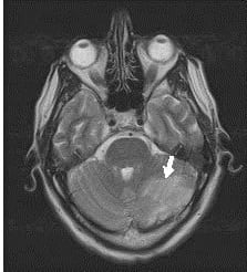
A 58-year-old male with a history of moderate hypertension and treatment with enalapril (20 mg/day) presented to our emergency department with acute headache in both the cervical and occipital regions of left side. About two hours later he developed asterixis of the upper left limb and gait instability. There was no history of alcohol or drug abuse, and no significant past medical or surgical history. History did not reveal symptoms consistent with depression and/or anxiety, and the patient was only taking enalapril. Neurological examination showed gait instability. Biochemical tests, including serum ammonia, ceruloplasmin, and hematological tests, were normal. Blood pressure was 140/80 mmHg and electrocardiogram showed a sinus rhythm (72 beats/min). Baseline electroencephalogram disclosed mild, generalized, background alpha activity. Ultrasound of the neck was normal. Computer tomography of brain did not reveal structural lesions or abnormalities. The hyperkinetic movement disorder diminished in intensity and was observed as a pattern of asterixis only when the hands were stretched. Over the next 3 days, the asterixis disappeared and neurological examination revealed a weakness in the upper left limb (IV/V on the 0–V Medical Research Council scale). Magnetic resonance imaging (MRI) of the brain disclosed cerebellar infarctions in the left superior cerebellar artery region (Figure). Because angio-MRI did not reveal abnormality, we started the patient on aspirin (100 mg/day). Five days later, clinical examination revealed a weakness in the upper left limb (IV/V) with slight gait instability and the patient was discharged. Clinical evaluations performed one month later did not reveal any signs of neurological lesions.
DISCUSSION
Asterixis may be induced by a focal structural brain lesion such as a stroke and involves the midbrain, thalamus, parietal lobe, or frontal cortex.1,2,5 The incidence of post-stroke asterixis remains unknown. Previous studies reported bilateral asterixis due to unilateral brain lesions after infarction in the anterior cerebral artery region.2,5 In our patient the asterixis started immediately after the onset of stroke at the superior cerebellar artery and resolved within 3 days. The pathogenic mechanism for asterixis remains elusive. Previously it has been reported that asterixis is a negative myoclonus caused by intermittent failure in maintaining sustained muscle contraction.1,2 Both the anatomic location and the presence of gait instability in our patient suggest that asterixis may reflect a failure in arm posture maintenance, inducing the failure in the leg posture control. The postural stability or tonic control of the extremities is related to multiple brainstem–spinal pathways, such as the vestibulospinal, reticulospinal, and rubrospinal tracts. These systems are regulated by supratentorial structures; the ventral lateral nucleus of the thalamus is the area where the cerebellar–rubral or vestibulocerebellar fibers converge, and it is also strongly connected with the prefrontal area.4 The projections from the medial frontal cortex to the brainstem reticular formation may play a role in the regulation of muscle tone or posture.4 The occasional occurrence of bilateral asterixis and the transient nature of symptoms suggest that the system regulating posture maintenance is not strictly unilateral.
In a clinical study the author reported that the occurrence of ipsilateral asterixis in patients with cerebellar lesions can be explained by crossing cerebellar rubral fibers at the superior cerebellar peduncle. 4 Our patient’s asterixis was ipsilateral to acute cerebellar infarction, confirming that finding.
In conclusion, we reported that a transitory unilateral asterixis associated with ipsilateral headache can represent an initial clinical manifestation of ipsilateral cerebellar infarcts in the region of the superior cerebellar artery territory.
Footnotes
Supervising Section Editor: Rick A. McPheeters, DO
Submission history: Submitted September 9, 2011; Revision received December 15, 2011; Accepted January 1, 2012.
Full text available through open access at http://escholarship.org/uc/uciem_westjem
DOI:10.5811/westjem.2012.1.6900
Address for Correspondence: Antonio Siniscalchi, MD, Department of Neurology, Annunziata Hospital, Via F. Migliori, 1, 87100 Cosenza, Italy
E-mail: anto.siniscalchi@libero.it
Conflicts of Interest: By the WestJEM article submission agreement, all authors are required to disclose all affiliations, funding sources, and financial or management relationships that could be perceived as potential sources of bias. The authors disclosed none.
REFERENCES
1. Handley A, Medcalf P, Hellier K, et al. Movement disorders after stroke. Age and Aging.2009;38:260–6.
2. Kim JS. Asterixis after unilateral stroke: Lesion location of 30 patients. Neurology.2001;56:533–6. [PubMed]
3. Kim JS. Involuntary Movements After Anterior Cerebral Artery Territory Infarction.Stroke. 2001;32:258–61. [PubMed]
4. Edlow JA, Newman-Toker DE, Savitz SI. Diagnosis and initial management of cerebellar infarction. Lancet Neurol. 2008;7:951–64. [PubMed]
5. Rio J, Montalban J, Pujadas F, et al. Asterixis associated with anatomic cerebral lesions: a study of 45 cases. Acta Neurol Scand. 1995;91:377–81. [PubMed]


