| Author | Affiliation |
|---|---|
| James J. Ham, MD | Madigan Army Medical Center, Department of Emergency Medicine, Tacoma, WA |
| Jason D. Heiner, MD | Madigan Army Medical Center, Department of Emergency Medicine, Tacoma, WA |
| Lynn EJ Gower, DO | Madigan Army Medical Center, Department of Emergency Medicine, Tacoma, WA |
| Joseph S. Litner, MD | Madigan Army Medical Center, Department of Emergency Medicine, Tacoma, WA |
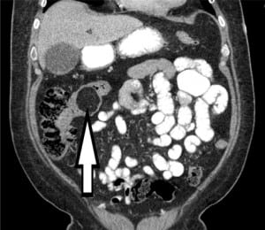
A 60-year-old male presented to the emergency department (ED) with 24 hours of progressive abdominal pain associated with nausea and vomiting. His physical exam revealed a soft but somewhat distended and tender abdomen, particularly over the mid abdomen and right upper quadrant. Plain radiography was concerning for bowel obstruction. Computed tomography revealed an ileocecal intussusception secondary to an intestinal lipoma as the cause of the obstruction (Figure). Diagnosis was confirmed in the operating room where he underwent laparotomy with excision of the mass via hemicolectomy.

Intestinal lipomas are rare, benign neoplasms, representing 2.6% of all non-malignant tumors of the intestinal tract and typically found on the right side of the colon.1,2 Colonic lipomas cause symptoms in less than 25% of patients, usually when the tumor grows to greater than 2 cm in diameter.2 These symptoms are typically vague, but lipomas have been known to cause bowel obstructions secondary to intussusceptions. Adult intussusception accounts for about two percent of bowel obstructions and constitutes approximately 5% of all intussusceptions.3 In contrast to children, more than 90% of adult intussusceptions have a demonstrable cause with 60% due to a neoplasm or other mass at least 5 cm in diameter.2 Colonic lipomas are often easily recognized on abdominal CT. Segmental colon resection is often required for symptomatic lipomas, particularly those greater than 2 cm and when malignancy cannot be ruled out.2
Footnotes
Supervising Section Editor: Sean Henderson, MD
Submission history: Submitted December 5, 2009; Accepted December 14, 2009
Full text available through open access at http://escholarship.org/uc/uciem_westjem
Address for Correspondence: James Ham, MD, Madigan Army Medical Center, Dept of Emergency Medicine, 9040 Fitzsimmons Ave, Tacoma, WA 98431
Email: james.ham@gmail.com
Conflicts of Interest: By the WestJEM article submission agreement, all authors are required to disclose all affiliations, funding sources, and financial or management relationships that could be perceived as potential sources of bias. The authors disclosed none.
REFERENCES
1. Siegal A, Witz M. Gastrointestinal lipoma and malignancies. J Surg Oncol. 1991;47:170–4.[PubMed]
2. Du L, Shah TR, Zenilman ME. Image of the month–quiz case. Intussuscepted transverse colonic lipoma. Arch Surg. 2007;142:1221. [PubMed]
3. Meshikhes AW, Al-Momen SA, Al Talaq FT, et al. Adult intussusception caused by a lipoma in the small bowel: report of a case. Surg Today. 2005;35:161–5. [PubMed]


