
| Author | Affiliation |
|---|---|
| Peter Fredericks, BS | University of Illinois-College of Medicine, Chicago, Illinois |
| Theresa Liu, BS | University of Illinois-College of Medicine, Chicago, Illinois |
| Joseph Colla, MD, RDMS | University of Illinois at Chicago, Department of Emergency Medicine, Chicago, Illinois |
CASE REPORT
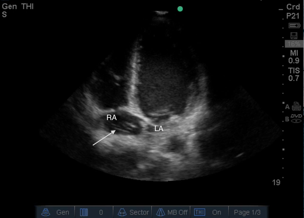
A 31-year-old African-American male with known sickle cell disease presented to the emergency department (ED) with a one-week history of chest pain and bilateral leg pain. Physical examination showed an afebrile and hemodynamically stable but uncomfortable appearing male. Cardiac and respiratory exam were unremarkable. A bedside focused cardiac ultrasound (FOCUS) exam performed by the attending emergency physician (EP) revealed four dilated chambers and a hyperechoic mobile body in the right atrium (Image). The FOCUS images were forwarded to a senior cardiology fellow who confirmed that the hyperechoic body was a Chiari network and not a right atrial clot. The patient was admitted for a sickle cell vaso-occlusive crisis that was managed with morphine and intravenous fluids.
CPC-EM Capsule
What do we already know about this clinical entity?
Chiari’s network is a collection of reticula found in the right atrium that has been associated with some cardiac pathologies such as arrhythmia, paradoxical emboli, and thrombi formation.
What is the major impact of image(s)?
The major impact of this image is to inform providers of this disease entity, emphasize its defining characteristics, and highlight the associated pathologies.
How might this improve emergency medicine practice?
A Chiari’s network can prove a diagnostic challenge for emergency physicians, at times mimicking a right atrial mass, thrombus, or vegetation which may in turn lead to mistreatment.
DISCUSSION
Found in 2% of the population, Chiari network is a collection of reticula in the right atrium that results from incomplete resorption of the Eustachian valve.1 The network is visualized on echocardiogram as a pulsating network of threads and fibers attached to the posterior wall of the right atrium or atrial septum. This sonographic appearance can prove to be a diagnostic challenge for EPs using point-of-care ultrasound because it could be mistaken for a right atrial mass, thrombus, or vegetation instead of an embryological remnant, which may in turn lead to mistreatment.2 Key elements noted on echocardiography that distinguish Chiari network include identification of at least two normal-appearing tricuspid valve leaflets and the presence of a rotary, highly mobile target that does not move into the right ventricular outflow tract or the right ventricle during diastole. While typically considered a benign anatomical variant, it has been associated with cardiac pathologies such as arrhythmia, paradoxical emboli, persistent patent foramen ovale, formation of an atrial septal aneurysm, thrombi formation, and entrapment of thrombi or catheters.1,3,4 Differentiating Chiari network from a more acutely pathological process is critical in the evaluation and management of hypercoagulable patients.
Footnotes
Section Editor: Rick A. McPheeters, DO
Full text available through open access at http://escholarship.org/uc/uciem_cpcem
Address for Correspondence: Peter Fredericks, BS, University of Illinois-College of Medicine, 808 South Wood St, Chicago, IL 60612. Email: peter.fredericks@gmail.com. 1:258 – 259
Submission history: Revision received October 10, 2016; Submitted February 14, 2017; Accepted February 22, 2017
Conflicts of Interest: By the CPC-EM article submission agreement, all authors are required to disclose all affiliations, funding sources and financial or management relationships that could be perceived as potential sources of bias. The authors disclosed none.
REFERENCES
1. Loukas M, Sullivan A, Tubbs RS, et al. Chiari’s network: review of the literature. Surg Radiol Anat. 2010;32(10):895-901.
2. Werner JA, Cheitlin MD, Gross BW, et al. Echocardiographic appearance of the Chiari network: differentiation from right-heart pathology. Circulation. 1981;63(5):1104-9.
3. Schneider B, Hofmann T, Justen MH, et al. Chiari’s network: normal anatomic variant or risk factor for arterial embolic events?. J Am Coll Cardiol. 1995;26(1):203-10.
4. Schwimmer-Okike N, Niebuhr J, Schramek G, et al. The presence of a large Chiari network in a patient with atrial fibrillation and stroke. Case Rep in Cardiol. 2016;2016:4839315.
SUPPLEMENTARY MATERIAL
An apical four chamber view displaying a prominent Chiari network within the right atrium.
An apical four chambier view displaying a right atrial thrombus to contrast with the Chiari network found in Video 1.