
| Author | Affiliation |
|---|---|
| Adam S. Bloom, DO | Naval Medical Center Portsmouth, Department of Emergency Medicine, Portsmouth, Virginia |
| Jonathan Auten, DO | Naval Medical Center Portsmouth, Department of Emergency Medicine, Portsmouth, Virginia |
| Joel M. Schofer, MD | Naval Medical Center Portsmouth, Department of Emergency Medicine, Portsmouth, Virginia |
CASE PRESENTATION
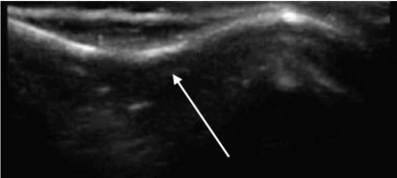
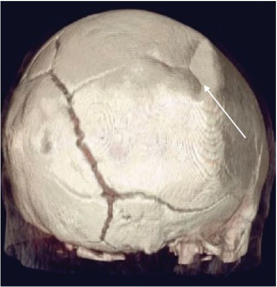
A four-month-old female presented to the emergency department after a witnessed fall from a high chair. She landed on her head but did not lose consciousness. She did not have any vomiting or altered mental status. There was a palpable defect in her right parietal skull. Point-of-care ultrasound (POCUS) demonstrated a large depression in her parietal skull consistent with a depressed skull fracture (Image 1). The fracture was confirmed by a non-contrast computed tomography (CT) of the head (Image 2). The CT was otherwise negative. The patient was admitted for observation but was discharged after an uncomplicated hospital course and was doing well at a follow-up visit.
CPC-EM Capsule
What do we already know about this clinical entity?
We know that ultrasound is a modality with excellent specificity for the diagnosis of skull fractures.
What is the major impact of the image(s)?
Ultrasound can also be used to diagnose depressed skull fractures in pediatric patients.
How might this improve emergency medicine practice?
Ultrasound may aid in the diagnosis of depressed skull fractures in pediatric patients.
DIAGNOSIS
Depressed skull fractures in neonates are typically different from those in adults. The soft bone tends to buckle rather than break. As such, they are often referred to as “ping pong” fractures, a reference to the way a ping pong ball looks when it has been indented. They may occur as sequelae to trauma, birth, or normal uterine development.1 POCUS is a convenient method to quickly and accurately detect skull fractures in pediatric patients with sensitivities ranging from 88–100% in two prospective trials.2-3 While most authors agree that a CT is indicated to rule out underlying pathology once a skull fracture is identified, a consensus does not exist on the role of POCUS to safely rule out skull fractures in neonates.4 We believe this is the first reported case of a “ping pong” skull fracture diagnosed using POCUS.
Footnotes
Section Editor: Rick A. McPheeters, DO
Full text available through open access at http://escholarship.org/uc/uciem_cpcem
Address for Correspondence: Adam S. Bloom, DO, Naval Medical Center Portsmouth, Department of Emergency Medicine, 620 John Paul Jones Circle, Portsmouth, VA 23708. Email: adambloom34@gmail.com. 2:99 – 100
Submission history: Revision received August 28, 2017; Submitted December 13, 2017; Accepted December 13, 2017
Conflicts of Interest: By the CPC-EM article submission agreement, all authors are required to disclose all affiliations, funding sources and financial or management relationships that could be perceived as potential sources of bias. The authors disclosed none. This work was prepared as part of authors’ official duties. Title 17 U.S.C. 105 provides that ‘Copyright protection under this title is not available for any work of the United States Government.’ Title 17 U.S.C. 101 defines a United States Government work as a work prepared by a military service member or employee of the United States Government as part of that person’s official duties. The views expressed in this article are those of the authors and do not necessarily reflect the official policy or position of the Department of the Navy, Department of Defense, or the United States Government.
REFERENCES
1. Zalatim O, Ranasinghe M, Dias M, et al. Treatment of depressed skull fractures in neonates using percutaneous microscrew elevation. J Neurosurg Pediatr. 2012;9(6):676-9.
2. Parri N, Crosby B, Glass C, et al. Ability of emergency ultrasonography to detect pediatric skull fractures: a prospective, observation study. J Emerg Med. 2013;44(1):135-41.
3. Rabiner J, Friedman L, Khine H, et al. Accuracy of point-of-care ultrasound for the diagnosis of skull fractures in children. Pediatrics. 2013;131:1757-64.
4. Ramirez-Schrempp D, Vinci RJ, Liteplo A. Bedside ultrasound in the diagnosis of skull fractures in the pediatric emergency department. Pediatr Emerg Care. 2011;27(4):312-4.