| Author | Affiliation |
|---|---|
| Asghar Haider, BS | University of California Irvine, School of Medicine, Irvine, California |
| Wirachin Hoonpongsimanont, MD | University of California Irvine, Department of Emergency Medicine, Orange, California |
ABSTRACT
Chest pain is a common presenting symptom in the emergency department. After ruling out emergent causes, emergency physicians need to identify and manage less commonly encountered conditions. Pulmonary sequestration (PS) is a rare congenital condition involving pulmonary parenchyma. In PS, a portion of non-functional lung tissue receives systemic blood supply from an anomalous artery. While most individuals with PS present in early life with symptoms of difficulty feeding, cyanosis, and dyspnea, some present later with recurrent pneumonia, hemoptysis, or productive cough. In this report, we present a case of PS in an adult with acute onset pleuritic chest pain.
CASE PRESENTATION
A previously healthy 26 year old male presented to the emergency department (ED) with chest pain for 1 hour. The patient stated the substernal pain was 10/10 in severity, non-radiating and worse with deep inspiration. He experienced intermittent left arm numbness that started after the onset of the chest pain but denied heart palpitations, diaphoresis, shortness of breath, hemoptysis, cough or recent fevers. On physical exam, vital signs were unremarkable, and he was cooperative and able to speak in full sentences. Chest auscultation did not detect any abnormalities. An electrocardiogram was read as normal. A chest radiograph revealed a lung lesion in the left lower lobe. A computed tomography (CT) of the chest showed a 11 cm × 9 cm × 10.5 cm round, well-defined, heterogeneous mass consistent in location with the lesion noted on the chest radiograph. The patient’s chest pain was well controlled on oral analgesics, and after consultation with the surgical service he was scheduled for an outpatient appointment for follow up and treatment. The patient was discharged and prescribed acetaminophen-hydrocodone 500 mg-5 mg oral tablets to be taken as needed for pain 1–2 tablets every 4–6 hours with a maximum of 8 tablets per day. He was advised to return to the ED if symptoms persisted or worsened.
The patient returned to the ED 1 day later with recurrent chest pain that was now more localized around his left lower ribs and rated as 8 out of 10 in severity. His vital signs remained within normal limits and his physical exam was unchanged from discharge. A repeat chest CT showed no changes. His pain management therapy was changed to acetaminophen-oxycodone 325 mg–7.5 mg oral tablets to be taken as needed for pain 1 tablet every 4 hours with a maximum of 6 tablets per day. This adjustment resulted in symptomatic relief of his chest pain. With improved symptoms and unchanged imaging, he was discharged with strict follow-up instructions.
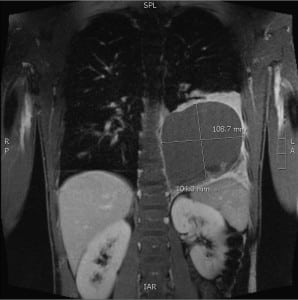
The patient returned 3 days later for his scheduled outpatient appointment. The surgical service obtained a chest magnetic resonance angiogram with contrast that revealed a cystic lesion measuring 108.7 mm by 104.0 mm (Figure). A thoracotomy with resection of an extralobar pulmonary sequestration (PS) was completed without intraoperative or post-operative complications. The non-communicating lung parenchyma was being supplied by an artery arising from the abdominal aorta. The patient was discharged home on post-operative day 4.

Chest magnetic resonance angiogram with contrast showing the extralobular pulmonary sequestration as a cystic lesion in the right lower lung field.
At a follow-up outpatient appointment 1 week after discharge, the patient remained asymptomatic, and a repeat chest radiograph showed resolution of the PS. The patient did not require further outpatient follow-up.
DISCUSSION
PS is a rare congenital pulmonary malformation involving lung parenchyma that lacks communication with the tracheobronchial tree. Arterial blood supply in most cases of PS is provided by the thoracic or abdominal aorta, and venous drainage is usually accomplished by pulmonary veins.1 Most cases are diagnosed and treated in childhood with diagnosis possible antenatally by ultrasound as early as 18–19 weeks of gestation.3 In the rare instances that PS presents in adulthood, symptoms like recurrent pneumonia, productive cough and hemoptysis are usually present.4 As our case demonstrates, patients may also present with chest pain and other nonspecific symptoms.
When PS is incidentally found in asymptomatic individuals, observation with close monitoring may be sufficient. Recently some authors have argued for resection in asymptomatic patients due to risk of infections, the low rate of natural regression, and to exclude other pathology.5
In cases of symptomatic patients, such as the one presented here, management strategy is directed at confirming diagnosis, alleviating symptoms and then removal of the abnormal lung tissue. Diagnosis involves the identification of the aberrant arterial vessel supplying the non-functional lung parenchyma. Since it is not possible to exclude malignant etiologies from PS based on imaging alone, surgical intervention and histological examination are required to confirm the diagnosis.7 Thus, PS requires a high index of suspicion once an aberrant artery is visualized on CT angiography. In the case presented, a repeat chest CT was ordered on the second ED admission due to worsening symptoms and the uncertainty of diagnosis. This was necessary to rule out progression or rupture of the lung lesion.
It is not possible to diagnose PS in adults based on physical exam and history alone. Although recurrent pneumonia may increase suspicion of PS, due to the rarity of the condition and variety of presenting symptoms, the task is formidable. In a retrospective study reviewing 2,625 PS cases from the Chinese National Knowledge Infrastructure, authors of the report were able to identify symptoms of cough or expectoration in over half of the cases followed by fever and hemoptysis.6 These non-specific findings highlight the difficulty a clinician faces when attempting to diagnose PS based on history and physical exam findings.
Surgical resection is favored as the most effective method of treatment. In certain cases an embolization of the aberrant artery preformed preoperatively can facilitate surgical resection.5 This technique is more valuable when removing intralobar PS, which are more challenging to distinguish compared to extralobar PS. In our case, embolization was not used or necessary in the resection of the extralobar PS.
Once life-threatening conditions and other etiologies of chest pain are eliminated, PS must remain on the differential diagnosis when lung lesions with anomalous arterial supply are seen on imaging. In the acute care setting, emergency physicians should tailor treatment for patients with PS with symptom relief until surgical resection can be preformed.
Footnotes
Address for Correspondence: Asghar Haider, BS. 7226 Palo Verde Rd, Irvine, CA 92697. Email: asgharh@uci.edu. 11 / 2013; 14:638 – 639
Submission history: Revision received March 20, 2013; Submitted June 3, 2013; Accepted July 10, 2013
Conflicts of Interest: By the WestJEM article submission agreement, all authors are required to disclose all affiliations, funding sources and financial or management relationships that could be perceived as potential sources of bias. The authors disclosed none.
REFERENCES
1. Cobett HJ, Humphrey GM Pulmonary sequestration. Paediatr Respir Rev. 2004; 5:59-68
2. Sade RM, Clouse M, Ellis FH The spectrum of pulmonary sequestration. Ann Thorac Surg. 1974; 18:644-658
3. Samuel M, Burge DM Management of antenatally diagnosed pulmonary sequestration associated with congenital cystic adenomatoid malformation. Thorax. 1999; 54:701-706
4. Montjoy C, Hadique S, Graeber G Intralobar bronchopulmonary sequestra in adults over age 50: case series and review. W V Med J. 2012; 108:8-13
5. Cho MJ, Kim DY, Kim SC Embolization versus surgical resection of pulmonary sequestration: Clinical experiences with a thoracoscopic approach. J Pediatr Surg. 2012; 47:2228-2233
6. Wei Y, Li F Pulmonary sequestration: a retrospective analysis of 2625 cases in China. Eur J Cardiothorac Surg. 2011; 40:e39-42
7. Gompelmann D, Eberhardt R, Heubel CP Lung sequestration: a rare cause for pulmonary symptoms in adulthood. Respir. 2011; 82:445-450


