
| Author | Affiliation |
|---|---|
| Kael Duprey, MD, JD | Maimonides Medical Center, Department of Emergency Medicine, Brooklyn, NY |
| Michelle Lin, MD | San Francisco General Hospital, Department of Emergency Medicine, San Francisco, CA |
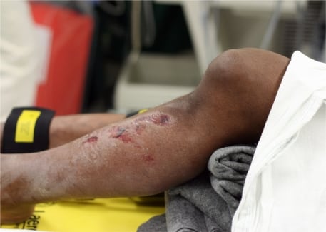
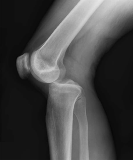
A 38-year-old male presented to the Emergency Department (ED) after a motorcycle crash. The patient was unable to walk because of isolated left knee pain. There were multiple abrasions over his left anterior tibia and a deformity of the left knee (Figure 1). The patient had very limited range of motion of his knee because of pain. His pedal pulses were normal bilaterally. The lateral view of the left knee showed a posterior knee dislocation (Figure 2). The patient’s knee was relocated in the ED, and serial ankle-brachial indices were monitored as an inpatient. No angiography was performed. An MRI showed significant ligamentous damage, including tears of the anterior and posterior cruciate ligament (ACL, PCL) and lateral collateral ligament (LCL). The medial collateral ligament (MCL) was intact. The patient received an external fixator of the knee in the operating room and was discharged home with close orthopedics follow up.
Knee dislocations are high-energy injuries. It is supported by the ACL and PCL, as well as the MCL and LCL. The disruption of all or most of these structures are required in knee dislocations. Complications include ligamentous and meniscal injuries, in addition to popliteal artery, popliteal vein, and peroneal nerve injuries. Concurrent popliteal artery injury has been reported to be 10–40%.1 Traditionally routine arteriography has been recommended for all patients with knee dislocations regardless of a normal distal vascular exam, because of the risk of an occult popliteal artery injury and thus potential risk for limb amputation. Recently this recommendation has been challenged.2 Early studies suggest Doppler ultrasonography and serial ankle-brachial indices may adequately rule-out arterial injury.3 Management is early knee relocation using longitudinal traction and prompt orthopedic referral. Arteriography should be performed for knee dislocations, suspicious for any popliteal arterial injury.
Footnotes
Supervising Section Editor: Sean Henderson, MD
Submission history: Submitted August 13, 2009; Revised August 14, 2009; Accepted August 28, 2009
Full text available through open access at http://escholarship.org/uc/uciem_westjem
Address for Correspondence: Michelle Lin, MD, 1001 Potrero Avenue, Suite 1E21, SFGH Emergency Medicine San Francisco, CA 94110
Email: michelle.lin@emergency.ucsf.edu
Conflicts of Interest: By the WestJEM article submission agreement, all authors are required to disclose all affiliations, funding sources, and financial or management relationships that could be perceived as potential sources of bias. The authors disclosed none.
REFERENCES
1. Stannard JP, Sheils TM, Lopez-Ben RR, et al. Vascular injuries in knee dislocations: the role of physical examination in determining the need for arteriography. J Bone Joint Surg Am. 2004;86-A:910–915. [PubMed]
2. Abou-Sayed H, Berger DL. Blunt lower-extremity trauma and popliteal artery injuries: revisiting the case for selective arteriography. Arch Surg. 2002;137:585–589. [PubMed]
3. Mills WJ, Barei DP, McNair P. The value of the ankle-brachial index for diagnosing arterial injury after knee dislocation: a prospective study. J Trauma. 2004;56:1261–5. [PubMed]